
Pulmonary tuberculosis detection method and system based on chest X-ray image and storage medium

A detection method and technology for pulmonary tuberculosis, applied in the field of medical image processing, can solve the problem of heavy workload of medical staff, and achieve the effect of reducing workload

Pending Publication Date: 2020-10-09
佛山市第四人民医院 +1
View PDF6 Cites 8 Cited by
  • Summary
  • Abstract
  • Description
  • Claims
  • Application Information

AI Technical Summary

Problems solved by technology

The current chest X-ray image processing method can only realize the extraction of the lung contour, which makes the workload of medical staff in the diagnosis of pulmonary tuberculosis larger

Method used


Image

  • Pulmonary tuberculosis detection method and system based on chest X-ray image and storage medium
  • Pulmonary tuberculosis detection method and system based on chest X-ray image and storage medium
  • Pulmonary tuberculosis detection method and system based on chest X-ray image and storage medium

Examples


Embodiment Construction

[0036] The idea, specific structure and technical effects of the present invention will be clearly and completely described below in conjunction with the embodiments and accompanying drawings, so as to fully understand the purpose, scheme and effect of the present invention.

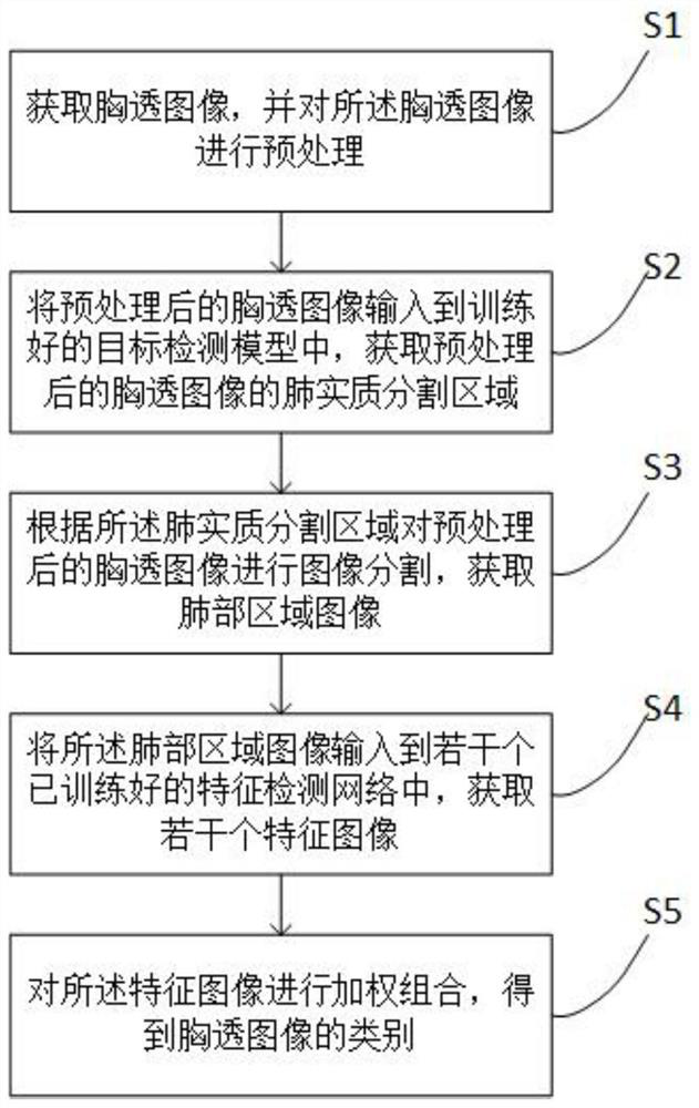
[0037] Embodiments of the present invention provide a method for detecting pulmonary tuberculosis based on chest X-ray images, referring to figure 1 , including the following steps:

[0038] S1. Obtain a chest X-ray image, and perform preprocessing on the chest X-ray image;

[0039] S2. Input the preprocessed chest X-ray image into the trained target detection model, and obtain the lung parenchyma segmentation area of ​​the preprocessed chest X-ray image;

[0040] S3. Perform image segmentation on the preprocessed chest X-ray image according to the lung parenchyma segmentation area, and obtain an image of the lung area;

[0041] S4. Input the images of the lung region into several trained feature detec...


PUM


Abstract

The invention discloses a pulmonary tuberculosis detection method and system based on a chest X-ray image and a storage medium, and the method comprises the following steps: obtaining the chest X-rayimage, and carrying out the preprocessing of the chest X-ray image; inputting the preprocessed chest X-ray image into a trained target detection model, and obtaining a lung parenchyma segmentation region of the preprocessed chest X-ray image; performing image segmentation on the preprocessed chest X-ray image according to the lung parenchyma segmentation region to obtain a lung region image; inputting the lung region image into a plurality of trained feature detection networks to obtain a plurality of feature images; and performing weighted combination on the feature image to obtain the category of the chest X-ray image. According to the invention, the chest radiography image is processed through the target detection model and the feature detection network, the pulmonary tuberculosis probability of the chest radiography image is predicted, and the workload of medical staff in pulmonary tuberculosis diagnosis is greatly reduced. The invention can be widely applied to the field of medical image processing.

Description

technical field [0001] The invention relates to the technical field of medical image processing, in particular to a method, system and storage medium for detecting pulmonary tuberculosis based on chest X-ray images. Background technique [0002] Tuberculosis is a chronic infectious disease transmitted through the respiratory tract. In recent years, due to multiple drug-resistant tuberculosis, double infection of tuberculosis and HIV, and the increase in floating population, the tuberculosis epidemic has rebounded. Tuberculosis has seriously endangered the health of the general public and has become a major public health and social problem. The key to doing a good job in the prevention and control of tuberculosis is to do a good job in the two important links of discovery and treatment and management of patients. Chest X-ray images play an important role in the initial screening of pulmonary tuberculosis. Pulmonary nodules have a wide variety of medical signs, with complex ...

Claims


Application Information

Patent Timeline
IPC IPC(8): G06T7/00G06T7/11G06K9/62G06N3/04G16H30/40
CPCG06T7/0012G06T7/11G16H30/40G06T2207/30064G06N3/045G06F18/214
Inventor 周杰邓东华龙显荣王威林东子霍颖瑜刘欣张浩洋
Owner 佛山市第四人民医院
Learn More