Visceral organ tissue high-identification imaging method and device, storage medium and computer equipment

An imaging method and tissue technology, applied in the field of medical image processing, can solve problems such as motion artifacts, misdiagnosis, and large loss, and achieve the effects of reducing radiation dose, high radiation dose, and low requirements

Pending Publication Date: 2020-11-20
广州海兆印丰信息科技有限公司
View PDF3 Cites 0 Cited by
  • Summary
  • Abstract
  • Description
  • Claims
  • Application Information

AI Technical Summary

Problems solved by technology

[0003] With the development of digital X-ray photography, digital photography Dual Energy Subtraction (Dual Energy Subtraction, DES) technology has realized the separation of bone and soft tissue in X-ray chest film; Subtract images, and reconstruct them into soft tissue images and bone tissue images by image subtraction; however, the image quality of dual-energy subtraction is easily affected by factors such as breathing, heartbeat, and movement between two exposures, and motion artifacts will occur. It is easy to lead to misdiagnosis; in addition, short-term alternating high and low energy X-ray beams have high requirements on the tube and large losses. Due to the need for special equipment, only a few hospitals use DES devices, and two high and low energy exposures will also increase the radiation dose to the patient

Method used


Image

  • Visceral organ tissue high-identification imaging method and device, storage medium and computer equipment
  • Visceral organ tissue high-identification imaging method and device, storage medium and computer equipment
  • Visceral organ tissue high-identification imaging method and device, storage medium and computer equipment

Examples


Embodiment

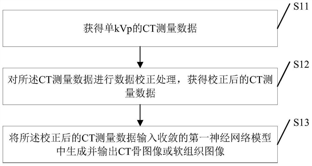
[0052] see figure 1 , figure 1 It is a schematic flow chart of an artificial intelligence-based high-resolution imaging method for organs and tissues in an embodiment of the present invention.

[0053] Such as figure 1 As shown, a high-resolution imaging method for organs and tissues based on artificial intelligence, the method includes:

[0054] S11: obtaining CT measurement data of a single kVp;

[0055] In the specific implementation process of the present invention, by obtaining the CT measurement data of the kVp (kilovolt peak value, which is the output capability unit of the CT equipment) of the interventional radiology equipment, that is, by setting the corresponding kVp data on the CT equipment, and then setting the corresponding kVp data in the kVp Set the corresponding CT measurement data on the basis of the data, and the CT measurement data of single kVp can be obtained.

[0056] S12: Perform data correction processing on the CT measurement data to obtain correc...


PUM


Abstract

The invention discloses an visceral organ tissue high-identification imaging method and device, a storage medium and computer equipment. The method comprises the steps: obtaining CT measurement data of a single kVp; performing data correction processing on the CT measurement data to obtain corrected CT measurement data; inputting the corrected CT measurement data into a converged first neural network model to generate and output a CT bone image or a soft tissue image. In the method disclosed in the embodiment of the invention, a high-quality CT bone image or soft tissue image can be obtained only by inputting conventional single-kVp CT measurement data, the efficiency is high, and the radiation dose of a patient is reduced.

Description

technical field [0001] The present invention relates to the technical field of medical image processing, in particular to an artificial intelligence-based high-resolution imaging method, device, storage medium and computer equipment for organs and tissues. Background technique [0002] Chest X-ray is an imaging technique widely used in the diagnosis of lung diseases because it is inexpensive, routinely effective, and relatively safe; however, the overlapping of anatomical structures such as ribs and clavicles makes it difficult for radiologists to read the diagnosis ; Therefore, the separation of bone structures on chest X-rays is of great help to radiologists and computer-aided diagnosis programs. [0003] With the development of digital X-ray photography, digital photography Dual Energy Subtraction (Dual Energy Subtraction, DES) technology has realized the separation of bone and soft tissue in X-ray chest film; Subtract images, and reconstruct them into soft tissue images...

Claims


Application Information

Patent Timeline
Patent Type & Authority Applications(China)
IPC IPC(8): G06T5/00G06T3/40G06T3/60G06N3/04
CPCG06T5/002G06T3/40G06T3/60G06T2207/10081G06T2207/30008G06N3/045
Inventor 谌高峰张浩蔡敏占
Owner 广州海兆印丰信息科技有限公司
Why Eureka
Try Eureka