
Melanoma segmentation method based on cavity convolution and multi-scale fusion

A multi-scale fusion, melanoma technology, applied in neural learning methods, image analysis, character and pattern recognition, etc., can solve the problems of information redundancy, blurred borders of melanoma regions, different shapes, etc., to suppress information redundancy. Effects of noise, mitigation of insufficient features, and reduction of semantic gaps

Pending Publication Date: 2021-03-05
ZHEJIANG UNIV OF TECH
View PDF0 Cites 11 Cited by
  • Summary
  • Abstract
  • Description
  • Claims
  • Application Information

AI Technical Summary

Problems solved by technology

Since the shallow features are not processed and directly integrated with the deep features, it will lead to information redundancy, which will affect the segmentation accuracy results.
[0004] Due to the fuzzy boundaries and various shapes of melanoma regions, it is difficult to accurately segment melanoma for general segmentation networks
There may be a strong connection between a large range of pixels in medical images. However, the general segmentation network usually uses a fixed-size convolution kernel to downsample the image, which causes the network to only capture local context information.
The proposed Atrous Space Convolution Pooling Pyramid (ASPP) can only extract part of the context information after downsampling, and cannot produce compact multi-scale features.

Method used


Image

  • Melanoma segmentation method based on cavity convolution and multi-scale fusion
  • Melanoma segmentation method based on cavity convolution and multi-scale fusion
  • Melanoma segmentation method based on cavity convolution and multi-scale fusion

Examples


Embodiment Construction

[0028] The present invention will be further described below in conjunction with accompanying drawing:

[0029] The melanoma segmentation method based on hole convolution and multi-scale fusion of the present invention comprises the following steps:

[0030] Step 1) preprocessing medical images;

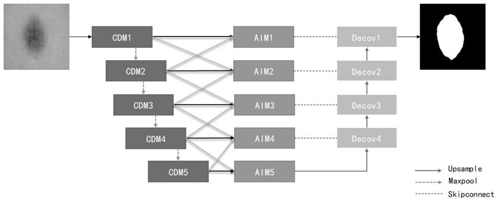
[0031] Divide the collected dermoscopic image data into training set, verification set and test set according to 7:1:2, and set the image pixel size to 128*128; do data augmentation on the training set images used for network training Processing, random rotation in the range of -30° to 30°, random horizontal flip and random scaling to between 0.8 and 1.2 times the original image;

[0032] Step 2) constructing a multi-scale aggregation network model with flexible receptive fields;

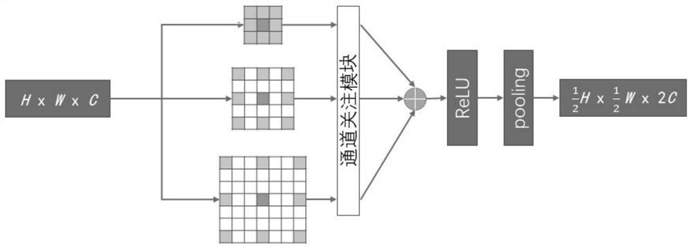
[0033] 2.1 Construct a channel attention hole convolution module for feature extraction;

[0034] Replace the encoding layer in U-Net with the channel attention hole convolution layer, take the dermosco...


PUM


Abstract

The invention discloses a melanoma segmentation method based on cavity convolution and multi-scale fusion. The melanoma segmentation method comprises the steps of 1) pre-processing a medical image; 2)constructing a multi-scale aggregation network model with a flexible receptive field; 3) inputting the training set data into the model for training; and 4) performing dermatoscope image lesion region segmentation. According to the channel attention hole convolution module provided by the invention, the receptive field can be adaptively expanded according to image features, tighter context information is obtained, and the problem of insufficient features caused by a fixed receptive field is relieved; an aggregation interaction module can aggregate the features output by a coding layer and thefeatures of an adjacent coding layer to obtain multi-scale information, reduce the semantic difference between the coding layer and the corresponding decoding layer, and suppress noise caused by direct aggregation. According to the method, a dermatoscope image can be segmented accurately, and an auxiliary effect is achieved.

Description

technical field [0001] The invention relates to a segmentation method for skin cancer melanoma Background technique [0002] Melanoma is one of the most dangerous skin diseases. Early studies have found that the 5-year survival rate for the most aggressive form of melanoma can be as high as 99%; however, delayed diagnosis can reduce survival to as low as 23%. Dermoscopy is the examination of skin lesions with a dermatoscope, often used to diagnose melanoma. However, manual inspection of dermoscopic images is error-prone and time-consuming, even for professional dermatologists. [0003] Therefore, it is necessary to develop a computational support system to assist dermatologists in accurately segmenting melanoma. This task remains challenging due to the fact that melanomas come in different sizes, shapes and textures. Also, some dermoscopic images may contain distractors such as hair, scale marks, and color calibration. Convolutional neural networks are heavily used to s...

Claims


Application Information

Patent Timeline
IPC IPC(8): G06T7/11G06T7/00G06T7/41G06K9/46G06K9/62G06N3/04G06N3/08
CPCG06T7/11G06T7/0012G06T7/41G06N3/08G06T2207/20081G06T2207/20084G06T2207/30096G06T2207/30088G06V10/462G06N3/045G06F18/253G06F18/214
Inventor 张聚潘伟栋俞伦端陈德臣牛彦施超
Owner ZHEJIANG UNIV OF TECH
Learn More