Tumor image brain region segmentation method and system based on image completion

An image and tumor technology, which is applied in the field of tumor image brain segmentation method and system, can solve the problems of strong subjectivity, high labor cost, and large gap between tumor parts, and achieve good segmentation effect, good adaptability, and improved accuracy Effect

Pending Publication Date: 2021-03-19
BEIJING ANDE YIZHI TECH CO LTD
View PDF5 Cites 8 Cited by
  • Summary
  • Abstract
  • Description
  • Claims
  • Application Information

AI Technical Summary

Problems solved by technology

[0005] The existing brain region segmentation methods for tumor images have the following disadvantages: 1) Manually label tumor brain regions: manual labeling of tumor brain regions faces the problems of high labor costs and strong subjectivity
2) Segmentation through the traditional brain partition model: this method has a poor segmentation result for the tumor part due to the large gap between the tumor image and the normal brain image in the tumor part
3) Segmentation by registration: The registration of brain tumor images has a large gap with normal brain images due to the existence of tumor parts, and the registration effect is poor, which in turn affects the segmentation effect
4) Training tumor image brain segmentation network: Due to the variety of tumor locations and shapes and the lack of tumor data, this method makes network learning tumor image brain segmentation difficult.

Method used


Image

  • Tumor image brain region segmentation method and system based on image completion
  • Tumor image brain region segmentation method and system based on image completion
  • Tumor image brain region segmentation method and system based on image completion

Examples


Embodiment Construction

[0061] The following will clearly and completely describe the technical solutions in the embodiments of the present invention with reference to the accompanying drawings in the embodiments of the present invention. Obviously, the described embodiments are only some, not all, embodiments of the present invention. Based on the embodiments of the present invention, all other embodiments obtained by persons of ordinary skill in the art without making creative efforts belong to the protection scope of the present invention.

[0062] In order to make the above objects, features and advantages of the present invention more comprehensible, the present invention will be further described in detail below in conjunction with the accompanying drawings and specific embodiments.

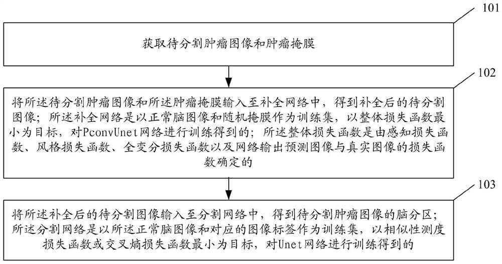
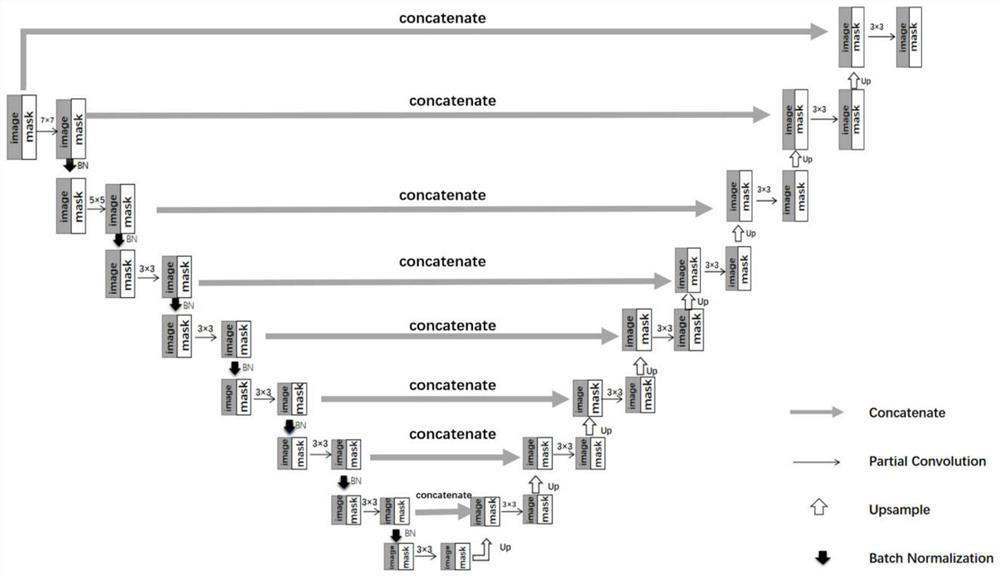
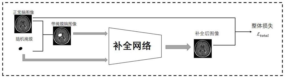
[0063] The tumor image brain region segmentation method based on image completion provided in this embodiment uses a partial convolution Unet network (PconvUnet network) to simultaneously update the tumor image to ...


PUM


Abstract

The invention discloses a tumor image brain region segmentation method and system based on image completion. The method comprises the steps of obtaining a to-be-segmented tumor image and a tumor mask;inputting the to-be-segmented tumor image and the tumor mask into a completion network to obtain a completed to-be-segmented image; wherein the completion network is obtained by training a PconvUnetnetwork by taking a normal brain image and a random mask as a training set and taking the minimum overall loss function as a target; inputting the completed to-be-segmented image into the segmentationnetwork to obtain brain partitions of the to-be-segmented tumor image; wherein the segmentation network is obtained by training the Unet network by taking a normal brain image and a corresponding image label as a training set and taking the minimum similarity measure loss function or the minimum cross entropy loss function as a target. The accuracy of brain region segmentation of the tumor imagecan be improved.

Description

technical field [0001] The present invention relates to the field of image segmentation, in particular to a method and system for brain region segmentation of tumor images based on image complementation. Background technique [0002] Brain tumors mostly grow in the cranial cavity, also known as intracranial tumors and brain cancers, which can originate from the brain, meninges, nerves, blood vessels, and brain appendages, or invade the brain from other tissues or organs of the body. The incidence of brain tumors is about 1.9 to 5.4 people / (year·100,000 people), accounting for 1% to 3% of all kinds of tumors in the whole body. Clinically, brain imaging data obtained by CT (Computed Tomography) and MRI (Magnetic Resonance Imaging) are mainly used for medical image processing. [0003] Due to the influence of image acquisition equipment, medical images (such as MRI images) will have problems such as low contrast, low signal-to-noise ratio, and low light intensity; and the orga...

Claims


Application Information

Patent Timeline
Patent Type & Authority Applications(China)
IPC IPC(8): G06T7/11G06T5/30G06N3/08G06T7/136G06N3/04G06K9/62
CPCG06T7/11G06T7/136G06T5/30G06N3/084G06T2207/30016G06T2207/30096G06N3/045G06F18/214
Inventor 程健倪莺珈吴振洲付鹤蒋景英刘涛
Owner BEIJING ANDE YIZHI TECH CO LTD
Why Eureka
Try Eureka