
External indentation positioning method for pleuroperitoneal fluid

A positioning method, chest and abdomen technology, applied in the direction of diagnosis, application, medical science, etc., to achieve the effect of precise positioning

Inactive Publication Date: 2021-03-26
长沙市第一医院
View PDF6 Cites 0 Cited by
  • Summary
  • Abstract
  • Description
  • Claims
  • Application Information

AI Technical Summary

Problems solved by technology

[0006] In view of the deficiencies in the prior art, the purpose of the present invention is to solve the problem of projection positioning of the body surface before puncture of pleural and abdominal fluid, and to develop a method for assisting ultrasonic positioning of pleural and abdominal fluid external indentation positioning

Method used


Image

  • External indentation positioning method for pleuroperitoneal fluid
  • External indentation positioning method for pleuroperitoneal fluid
  • External indentation positioning method for pleuroperitoneal fluid

Examples


Embodiment Construction

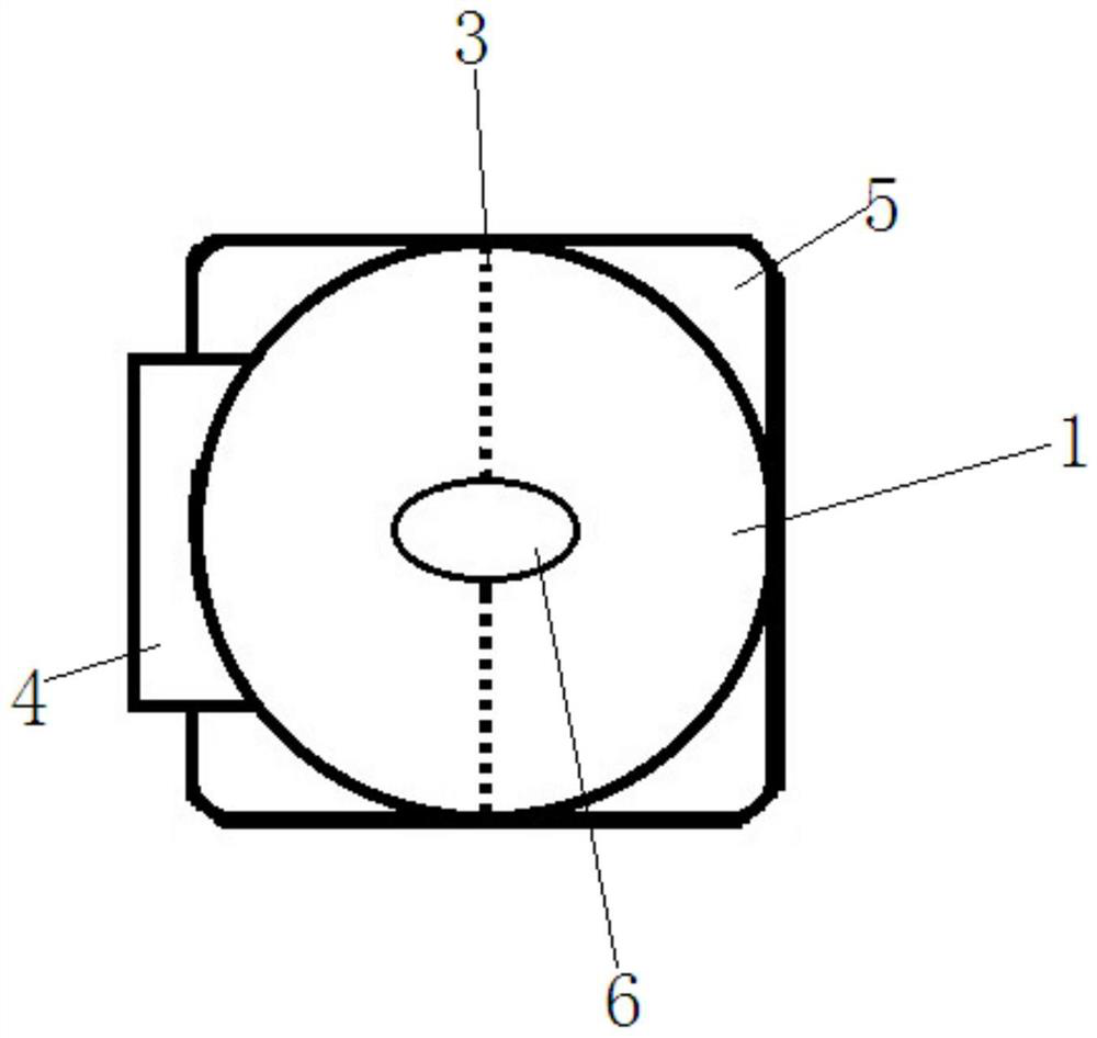
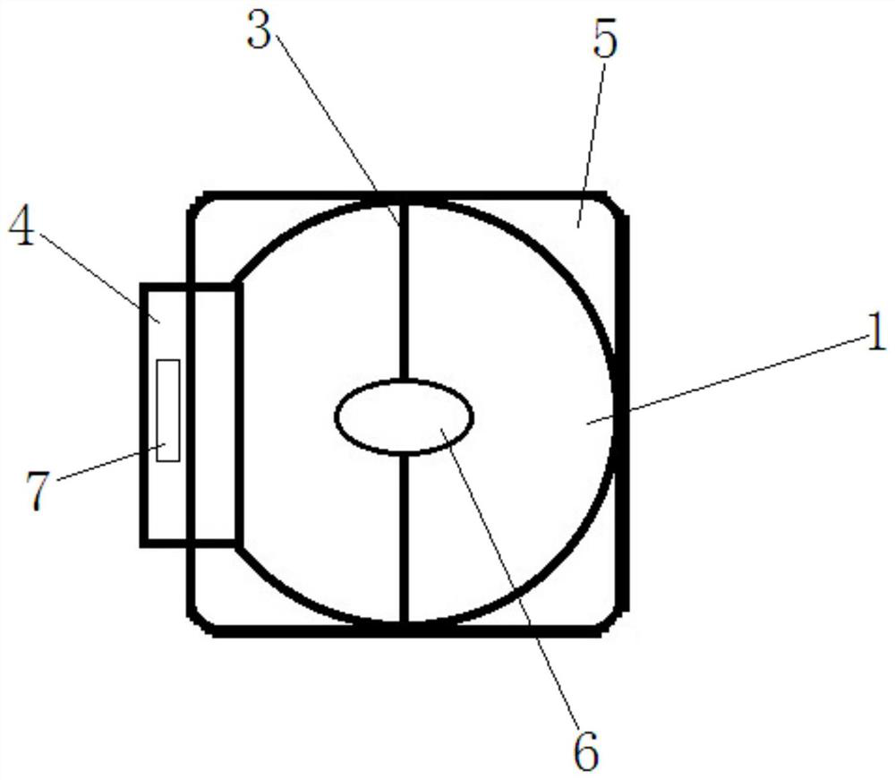
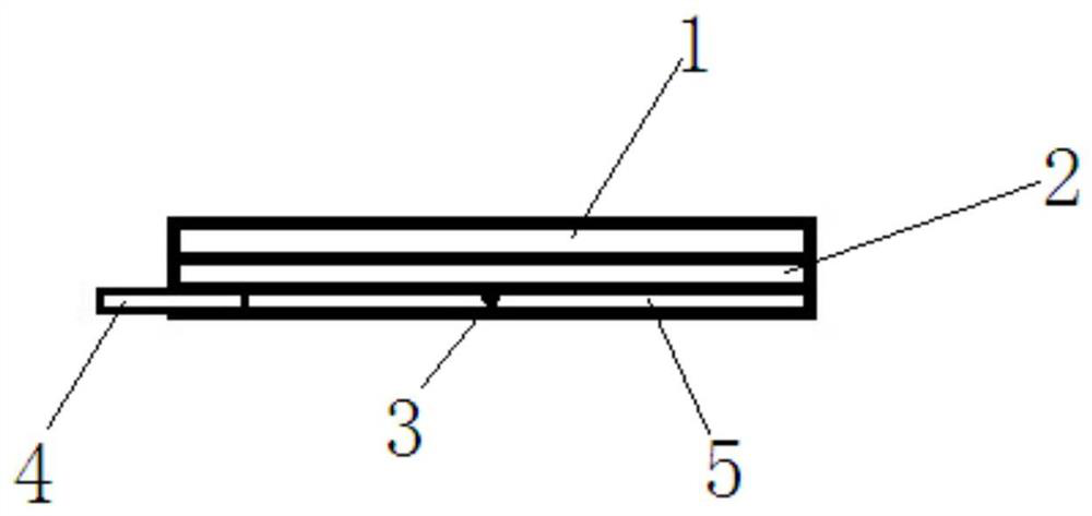
[0038] The detailed structure of the present invention will be further described below in conjunction with the accompanying drawings and specific embodiments.

[0039] A method for indentation positioning in vitro of thoracic and abdominal fluids, using indentation positioning stickers in vitro (such as Figure 1-6 As shown), the indentation positioning sticker for thoracic and abdominal water in vitro includes a waterproof and breathable back layer 1, an adhesive layer 2 and a release film 5 connected in sequence from top to bottom, and the adhesive layer 2 and the release film 5 are provided with Imprinting rod 3, the imprinting rod 3 is arranged symmetrically with the positioning through hole 6 as the center;

[0040] Also includes the following steps:

[0041] Step 1: Use ultrasound to detect the patient's pleural and ascites;

[0042] Step 2: Put the waterproof and breathable backing layer 1 of the external indentation positioning sticker on the thoracic and abdominal f...


PUM


Abstract

The invention provides an external indentation positioning method for pleuroperitoneal fluid, which adopts an external indentation positioning patch for pleuroperitoneal fluid, the indentation positioning patch is attached to the skin surface, pleuroperitoneal fluid can be accurately positioned through the indentation positions of two imprinting rods. The positioning method provided by the invention is simple and practical, the positioning is noninvasive, will not bring any pain to a patient, and can avoid cross infection effectively.

Description

technical field [0001] The invention relates to a device for locating the surface of human tissue, in particular to a tool for assisting ultrasound in locating pleural and ascites. Background technique [0002] Pleural and ascites, also known as pleural and peritoneal effusion, is one of the common complications of advanced cancer, and it is also the main clinical symptom or sign of some patients. Severe pleural and ascites can even be life-threatening. Pathological biopsy is an important method to confirm the diagnosis. Accurate localization is the key to effective pathological diagnosis. Treatment methods such as pumping and releasing water, etc., effectively controlling and eliminating cancerous pleural and ascites have important practical significance for prolonging and saving the lives of patients. [0003] Ultrasonic positioning of pleural and ascites before operation improves the accuracy of pleural and ascites puncture. At present, the location of pleural and asci...

Claims


Application Information

Patent Timeline
Patent Type & Authority Applications(China)
IPC IPC(8): A61B90/00A61B8/08A61B8/00
CPCA61B8/085A61B8/4444A61B90/39A61B2090/3904
Inventor 谭荣
Owner 长沙市第一医院
Learn More