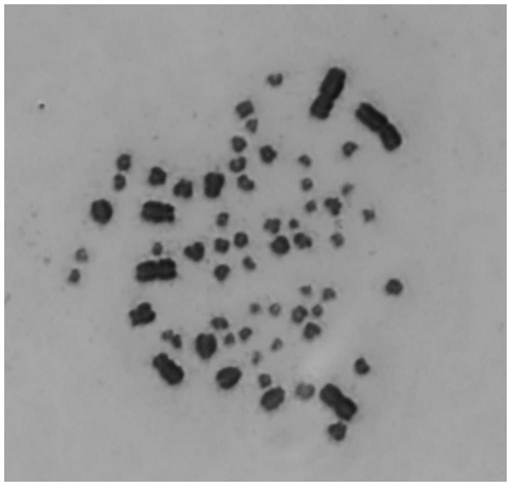
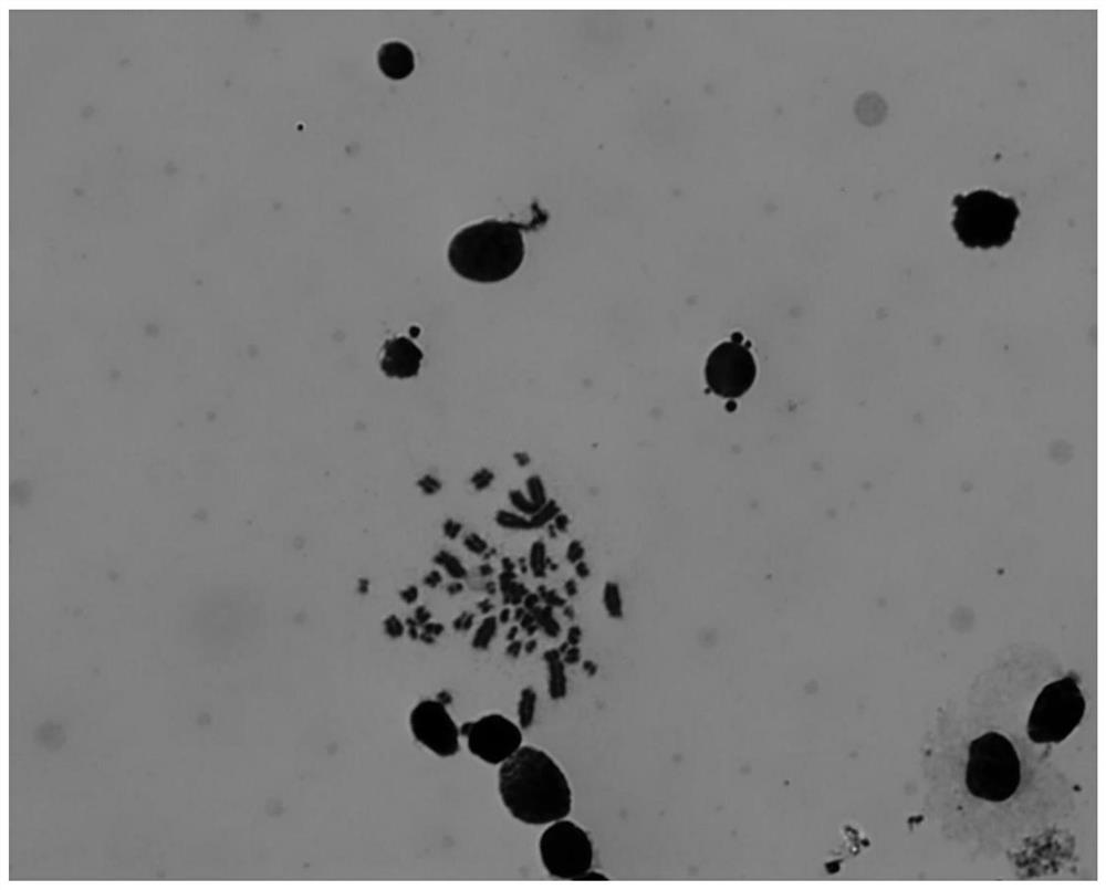
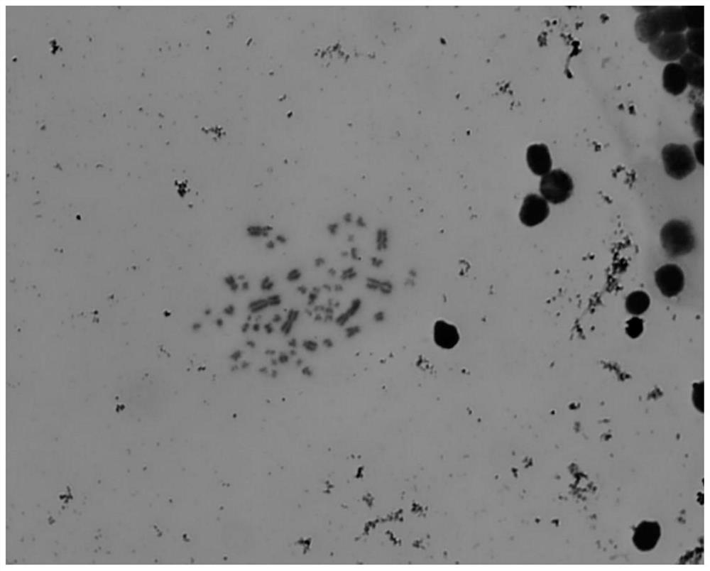
Method for preparing pelochelys biloba chromosome specimen by using peripheral blood cells
A chromosome and blood cell technology, applied in the field of cytogenetics, can solve the problems of scarcity and difficulty, and achieve the effect of easy observation and simple and fast production method
- Summary
- Abstract
- Description
- Claims
- Application Information
AI Technical Summary
Problems solved by technology
Method used
Image
Examples
Embodiment 1
[0033] 1. Peripheral blood sampling
[0034] Using a commercially available negative pressure blood collection tube (containing sodium heparin) and a sterilized needle, collect 1 ml of peripheral blood from the cervical sinusoids of the live C. After blood collection, it was temporarily stored at 4°C.
[0035] 2. Peripheral blood cell culture
[0036] Cell culture operations were performed under sterile conditions using a modified commercially available Eagle basal medium. The medium modified formula was added on the basal medium, 20mM 4-hydroxyethylpiperazineethanesulfonic acid, 1mM glutamate, 2nM sodium selenite, 1mM non-essential amino acids, 1mM sodium pyruvate, 1mM penicillin and strepto Mycin double antibody, 55μM β-mercaptoethanol, 15% fetal bovine serum, 5ng / mL recombinant human basic fibroblast growth factor. Add 200 μL of whole blood per 3 ml of medium, and add 750 μg of PHA-P.
[0037] 3. Colchicine treatment
[0038] Blood cells were cultured at 28°C (CO 2 Th...
Embodiment 2
[0042] 1. Peripheral blood sampling
[0043] Using a commercially available negative pressure blood collection tube (containing sodium heparin) and a sterilized needle, collect 1 ml of peripheral blood from the cervical sinusoids of the live C. After blood collection, it was temporarily stored at 4°C.
[0044] 2. Peripheral blood cell culture
[0045] Cell culture operations were performed under sterile conditions using a modified commercially available Eagle basal medium. The medium modified formula was added on the basal medium, 20mM 4-hydroxyethylpiperazineethanesulfonic acid, 1mM glutamate, 2nM sodium selenite, 1mM non-essential amino acids, 1mM sodium pyruvate, 1mM penicillin and strepto Mycin double antibody, 55μM β-mercaptoethanol, 15% fetal bovine serum, 5ng / mL recombinant human basic fibroblast growth factor. Add 200 μL of whole blood per 3 ml of medium, and add 750 μg of PHA-P.
[0046] 3. Colchicine treatment
[0047] Blood cells were cultured at 28°C (CO 2 The ...
Embodiment 3
[0051] 1. Peripheral blood sampling
[0052] Using a commercially available negative pressure blood collection tube (containing sodium heparin) and a sterilized needle, collect 1 ml of peripheral blood from the cervical sinusoids of the live C. After blood collection, it was temporarily stored at 4°C.
[0053] 2. Peripheral blood cell culture
[0054] Cell culture operations were performed under sterile conditions using a modified commercially available Eagle basal medium. The medium modified formula was added on the basal medium, 20mM 4-hydroxyethylpiperazineethanesulfonic acid, 1mM glutamate, 2nM sodium selenite, 1mM non-essential amino acids, 1mM sodium pyruvate, 1mM penicillin and strepto Mycin double antibody, 55μM β-mercaptoethanol, 15% fetal bovine serum, 5ng / mL recombinant human basic fibroblast growth factor. Add 200 μL of whole blood per 3 ml of medium, and add 750 μg of PHA-P.
[0055] 3. Colchicine treatment
[0056] Blood cells were cultured at 28°C (CO 2 Th...
PUM
 Abstract
Description
Claims
Application Information
 - R&D
- Intellectual Property
- Life Sciences
- Materials
- Tech Scout
- Unparalleled Data Quality
- Higher Quality Content
- 60% Fewer Hallucinations
Browse by: Latest US Patents, China's latest patents, Technical Efficacy Thesaurus, Application Domain, Technology Topic, Popular Technical Reports.
© 2025 PatSnap. All rights reserved.Legal|Privacy policy|Modern Slavery Act Transparency Statement|Sitemap|About US| Contact US: help@patsnap.com


