Electrocardiosignal premature beat detection method for

A technology for electrocardiographic signals and detection methods, which is applied in the directions of diagnostic recording/measurement, medical science, diagnosis, etc., and can solve the problems of poor detection effect of premature beats, noise interference, and poor detection effect.

Active Publication Date: 2021-04-09
SUZHOU UNIV
View PDF30 Cites 6 Cited by
  • Summary
  • Abstract
  • Description
  • Claims
  • Application Information

AI Technical Summary

Problems solved by technology

[0010] 1. For the noisy ECG signal, the obtained information such as the width and amplitude of the characteristic waveform is greatly disturbed by noise, and it is difficult to be used to detect premature beats
[0011] 2. This method relies on the characteristic waveform detection algorithm. If the characteristic waveform detection is not good, the subsequent detection effect will also deteriorate.
[0019] Most of the existing ECG signal premature beat detection algorithms rely on preprocessing work in the early stage, such as: heart beat detection, characteristic waveform detection, etc. If the preprocessing work is not handled well, the detection effect of premature beat beats in the later stage will also deteriorate
[0020] The existing ECG signal premature beat detection algorithm requires manual design of features: (1) Additional feature extraction and selection algorithms lead to an increase in computational complexity; (2) The quality of feature design directly affects the accuracy of feature wave detection; (3) When using fixed artificially designed features, it is difficult to maintain generalization ability;
[0021] The existing ECG signal premature beat detection algorithm is seriously affected by noise interference, and it is difficult to accurately locate the premature beat in the noisy ECG signal
[0022] The existing ECG signal premature beat detection algorithm steps are relatively cumbersome, unable to achieve end-to-end, fast premature beat detection, and the detection time is slow

Method used


Image

  • Electrocardiosignal premature beat detection method for
  • Electrocardiosignal premature beat detection method for
  • Electrocardiosignal premature beat detection method for

Examples


Embodiment Construction

[0048] The present invention will be further described below in conjunction with the accompanying drawings and specific embodiments, so that those skilled in the art can better understand the present invention and implement it, but the examples given are not intended to limit the present invention.

[0049] A method for detecting premature beats of electrocardiographic signals proposed by the present invention has achieved good results in the detection of premature beats of electrocardiographic signals. The complete technical solution is as follows:

[0050] S1. Data preparation

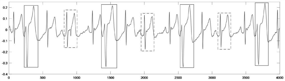
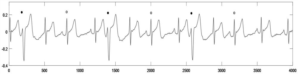
[0051] 1. the electrocardiographic signal data sampling rate that the present invention prepares is 400HZ, and the input length of electrocardiographic signal is fixed as uniform length (time length is 10 seconds, 4000 points), and electrocardiographic signal has electrode interference noise, myoelectricity in various degrees Interference noise and baseline drift noise.

[0052] 2. Prepare the corre...


PUM


Abstract

The invention discloses an electrocardiosignal premature beat detection method. The method comprises the following steps of step 1, fixing the input length of an electrocardiosignal with a preset sampling rate to be a uniform length, adding noise of different degrees to the electrocardiosignal, preparing a corresponding label for each group of data of the electrocardiosignal, providing the label with two channels, and dividing each channel into a corresponding ventricular premature beat QRS waveband and an supraventricular premature beat QRS waveband; and step 2, performing low-pass filtering on the electrocardiosignal in the step 1, inputting the electrocardiosignal into a neural network for training in combination with the corresponding label, a loss function of the neural network being a Dice loss function, and the neural network adopting a one-dimensional U-net network structure. The method has the beneficial effects that complex early-stage denoising or domain transformation is not needed, and manual feature design is not needed; the robustness is high, and the premature beat, namely the QRS wave position in the premature beat, can be accurately detected in a high-noise signal; and end-to-end detection is carried out, and a premature beat type and position are directly output through post-processing.

Description

technical field [0001] The invention relates to the field of electrocardiographic signal detection, in particular to a method for detecting premature beats of electrocardiographic signals. Background technique [0002] Electrodes are placed on different parts of the human body and connected to the positive and negative terminals of the electrocardiograph through lead wires. This circuit connection method for recording electrocardiograms is called electrocardiogram leads. The electrocardiogram is essentially a time-voltage graph of potential changes during heartbeat. In a normal cardiac cycle, a typical ECG waveform consists of a P wave, a QRS complex, a T wave, and a U wave that may be seen in 50% to 75% of ECGs. P waves correspond to atrial depolarization, QRS complexes correspond to ventricular depolarization, and T waves correspond to ventricular repolarization. Such as figure 1 Shown (refer to the national standard YY 0782-2010 / IEC60601-2-51: 2003). At present, the c...

Claims


Application Information

Patent Timeline
Patent Type & Authority Applications(China)
IPC IPC(8): A61B5/318A61B5/366A61B5/00
CPCA61B5/7203A61B5/7225A61B5/7267
Inventor 王丽荣蔡文强邱励燊朱文亮俞杰张淼王朵朵张慧敏
Owner SUZHOU UNIV
Why Eureka
Try Eureka