Image and depth-based cornea level identification and lesion positioning method and system

A positioning method and corneal technology, applied in the field of medical artificial intelligence image recognition, can solve the problems of incapable of intelligent positioning of lesions and insufficient accuracy of corneal level recognition, and achieve the effects of reducing invalid calculations, improving the accuracy of lesion recognition, and stabilizing the effect

Pending Publication Date: 2021-04-09
THE PEOPLES HOSPITAL OF GUANGXI ZHUANG AUTONOMOUS REGION
View PDF0 Cites 3 Cited by
  • Summary
  • Abstract
  • Description
  • Claims
  • Application Information

AI Technical Summary

Problems solved by technology

[0004] In order to overcome the existing problems of insufficient accuracy of corneal layer recognition and the inability to intelligently locate lesions, the present invention provides a method and system for corneal layer recognition and lesion location based on images and depths

Method used


Image

  • Image and depth-based cornea level identification and lesion positioning method and system
  • Image and depth-based cornea level identification and lesion positioning method and system
  • Image and depth-based cornea level identification and lesion positioning method and system

Examples


Embodiment Construction

[0035] In order to make the purpose, technical solutions and advantages of the present invention more clear, the present invention will be further described in detail below in conjunction with the accompanying drawings and implementation examples. It should be understood that the specific embodiments described here are only used to explain the present invention, not to limit the present invention.

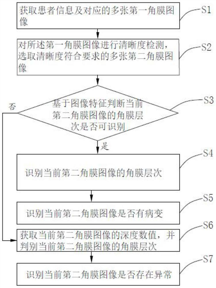
[0036] see figure 1 , the first embodiment of the present invention provides a corneal layer recognition and lesion location method based on images and depths, which includes the following steps:

[0037] Step S1: Acquiring patient information and multiple corresponding first corneal images;

[0038] Step S2: performing a sharpness detection on the first corneal image, and selecting a plurality of second corneal images whose sharpness meets the requirements;

[0039] Step S3: Determine whether the corneal layer of the current second corneal image is identifiable based on the imag...


PUM


Abstract

The invention relates to the field of medical artificial intelligence image identification, and particularly discloses an image and depth-based corneal hierarchy identification and lesion positioning method and system. After a corneal hierarchy image is obtained, image identification is carried out through a deep learning algorithm, the depth numerical analysis is carried out by combining with a machine learning algorithm, an anatomical level is automatically detected in cornea living body scanning, a level of a lesion or abnormal area can be accurately identified, visual reconstruction is performed on a lesion depth range, full-automatic level labeling and lesion positioning are realized, manual intervention is not needed, the labor cost is reduced, and meanwhile, corneal hierarchies are identified by using an analysis method integrated by multiple machine learning algorithms, the accuracy is high, and the effect is stable.

Description

technical field [0001] The invention relates to the field of medical artificial intelligence image recognition, in particular to a corneal layer recognition and lesion location method and system based on images and depths. Background technique [0002] Corneal disease can seriously threaten vision and is the second leading cause of blindness and low vision in my country. Confocal microscopy can scan the living cornea, detect the ultrastructure of the cornea, display the morphological changes at the cell level under normal and pathological conditions, and provide important information for the diagnosis of corneal diseases. By identifying the lesions and analyzing the anatomical layers involved, the severity of corneal disease can be assessed and an appropriate treatment plan can be selected. According to the anatomical structure and confocal microscope image morphology, the cornea can be divided into five layers: epithelial cell layer, Bowman's membrane, corneal stroma, Bowm...

Claims


Application Information

Patent Timeline
Patent Type & Authority Applications(China)
IPC IPC(8): G06T7/00G06K9/62
CPCG06T7/0012G06T2207/30041G06T2207/30096G06T2207/20081G06T2207/20084G06T2207/10028G06V10/751G06V2201/03
Inventor 徐帆唐宁宁蒋莉唐芬吕健黄光怡何文静
Owner THE PEOPLES HOSPITAL OF GUANGXI ZHUANG AUTONOMOUS REGION
Why Eureka
Try Eureka