Ultrasonic equipment, ultrasonic image processing method and storage medium

An ultrasound image and ultrasound equipment technology, which is applied in ultrasound/sonic/infrasound image/data processing, ultrasound/sonic/infrasonic Permian technology, organ motion/change detection, etc. Time repeated comparison and other issues to achieve the effect of improving accuracy, rapid determination, and real-time processing

Active Publication Date: 2021-04-16
QINGDAO HISENSE MEDICAL EQUIP
View PDF19 Cites 1 Cited by
  • Summary
  • Abstract
  • Description
  • Claims
  • Application Information

AI Technical Summary

Problems solved by technology

[0003] At present, there are two main methods for judging the end-systole and end-diastole of the left ventricle. One of the judgment methods is the manual observation method, that is, the doctor manually selects the frames of the end-systole and end-diastole images from the echocardiogram, and manually observes them. The method is relatively simple and intuitive, but it is subject to greater subjective influence, and it takes a lot of time to compare images repeatedly
Another judgment method is the electrocardiogram-assisted method, that is, when the doctor observes the echocardiogram, he combines the electrocardiogram to determine the left ventricle at the end of systole and the left ventricle at the end of diastole. Observe the inner diameter of the ventricle and the closure of the mitral valve by echocardiography, and subjectively determine the left ventricle at the end of systole and the left ventricle at the end of diastole

Method used


Image

  • Ultrasonic equipment, ultrasonic image processing method and storage medium
  • Ultrasonic equipment, ultrasonic image processing method and storage medium
  • Ultrasonic equipment, ultrasonic image processing method and storage medium

Examples


Embodiment Construction

[0077] In order to make the object, technical solution and advantages of the present invention clearer, the present invention will be further described in detail below in conjunction with the accompanying drawings. Obviously, the described embodiments are only some embodiments of the present invention, rather than all embodiments . Based on the embodiments of the present invention, all other embodiments obtained by persons of ordinary skill in the art without making creative efforts belong to the protection scope of the present invention.

[0078] The following is an explanation of some words that appear in the text:

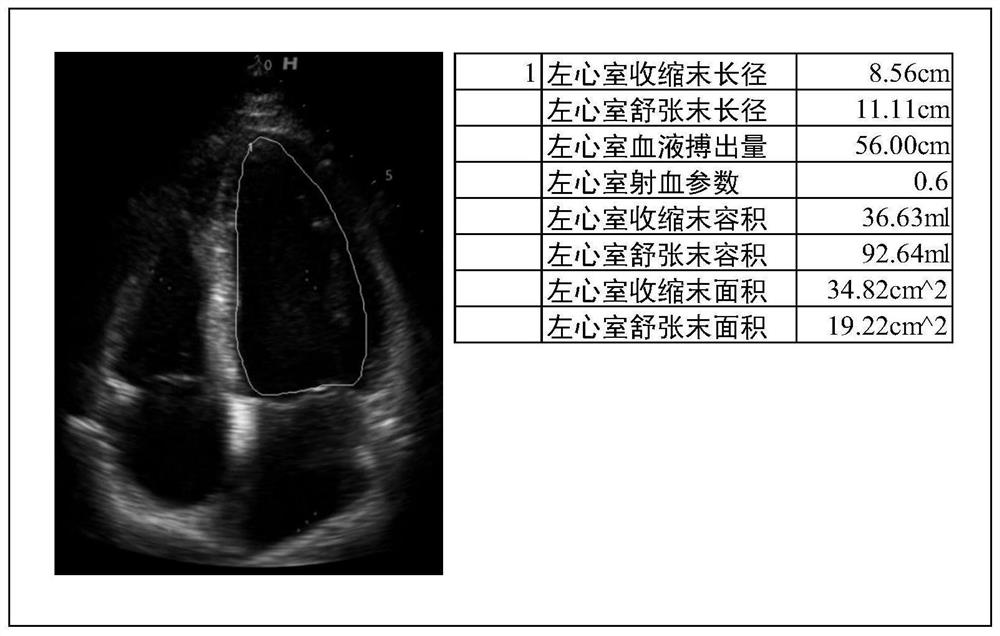
[0079] (1) Ejection parameter: refers to the percentage of blood stroke volume in the end-diastolic volume of the ventricle (ie, cardiac preload), the normal value is 50-70%, which can be checked by cardiac color Doppler ultrasound, which is an important factor in judging the type of heart failure One of the indications.

[0080] (2) Blood stroke volume: strok...


PUM


Abstract

The invention discloses ultrasonic equipment, an ultrasonic image processing method and a storage medium, which are used for automatically measuring heart state parameters and improving the measurement accuracy. The equipment comprises a processor and a display. The processor obtains an image sequence composed of ultrasonic images from the continuously collected ultrasonic image information of the heart; the ventricular volume corresponding to each frame of ultrasonic image in the image sequence is determined to obtain a ventricular volume sequence; the end-diastolic volume corresponding to the central ventricular end diastolic phase of at least one cardiac cycle and the end-systolic volume corresponding to the ventricular end-systolic phase is determined according to the change trend of the central ventricular volume of the ventricular volume sequence; and heart state parameters can be determined according to the end-diastolic volume and the end-systolic volume. The ventricular volume is determined according to the ultrasonic images in the acquired image sequence, so that the ventricular volume is determined quickly and accurately; the heart state parameters are determined according to the determined end-diastolic volume and end-systolic volume, so that the accuracy of the heart state parameters is improved.

Description

technical field [0001] The invention relates to the field of ultrasonic image processing, in particular to an ultrasonic device, an ultrasonic image processing method and a storage medium. Background technique [0002] Echocardiography can be used to monitor cardiac function and has the advantages of non-invasive and real-time observation. Ejection parameters refer to the percentage of the stroke volume of blood in the end-diastolic volume of the ventricle, which reflects the systolic function of the heart. Abnormal ejection parameters may be a sign of heart failure. The most commonly used method for measuring ejection parameters is echocardiography. Pulsed ultrasound passes through the chest wall and soft tissue to measure the periodic activities of the underlying heart walls, ventricles, and valves. volume, to calculate ejection parameters. [0003] At present, there are mainly two methods for judging the end-systole and end-diastole of the left ventricle. One of the ju...

Claims


Application Information

Patent Timeline
Patent Type & Authority Applications(China)
IPC IPC(8): A61B8/08
Inventor 张一琰时俊楠田广野吴乙荣王琦陈哲
Owner QINGDAO HISENSE MEDICAL EQUIP
Why Eureka
Try Eureka