
Infant intracranial hemorrhage fixing and guiding drainage device and preparation and use methods

A technology for intracranial hemorrhage and infants, which is applied in the field of medical equipment, can solve the problems of falling off, failed puncture drainage tube, inaccurate positioning of the body surface, etc., and achieves the effects of avoiding prolapse, improving accuracy, and avoiding unstable body position.

Pending Publication Date: 2021-05-14
QINGDAO WOMEN & CHILDREN HOSPITAL
View PDF0 Cites 1 Cited by
  • Summary
  • Abstract
  • Description
  • Claims
  • Application Information

AI Technical Summary

Problems solved by technology

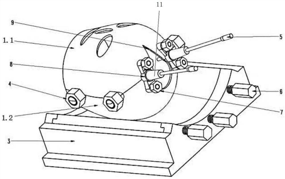
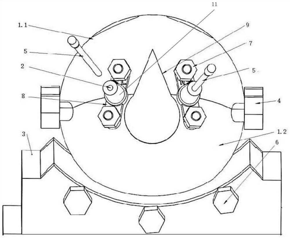
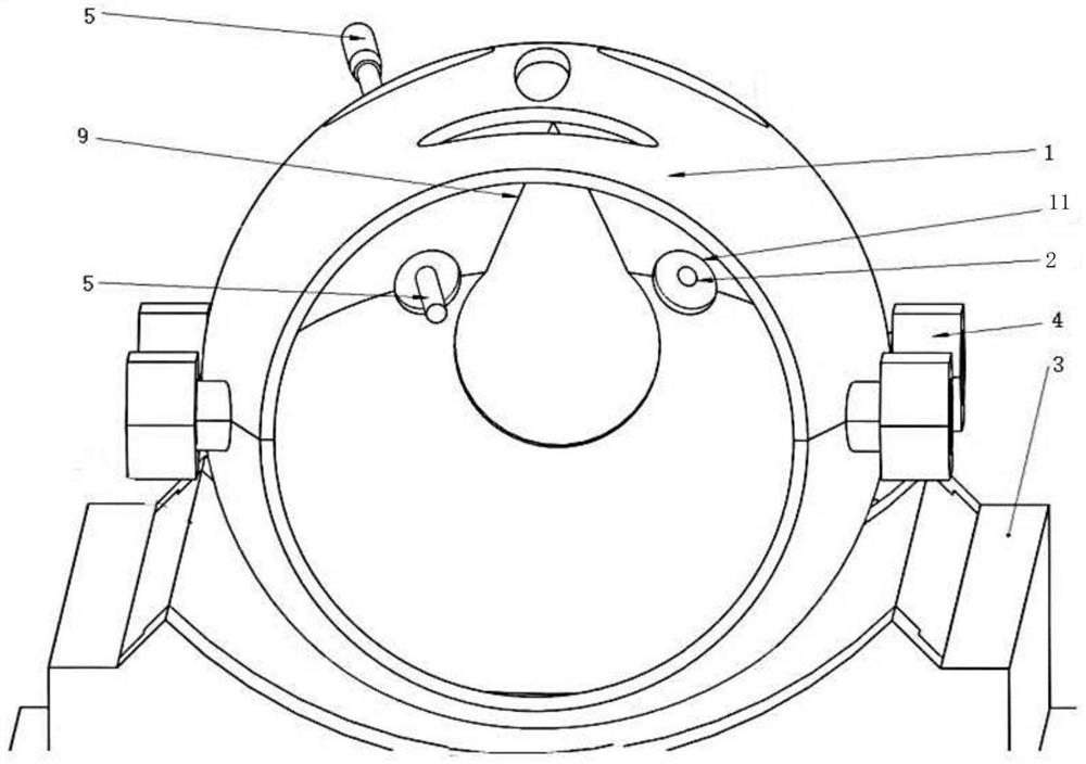
[0003] The technical problem to be solved by the present invention is to provide a fixed guiding and drainage device for intracranial hemorrhage in infants and its preparation and use method in view of the deficiencies in the prior art, so as to solve the problems of low puncture accuracy and poor body surface positioning in children with cerebral hemorrhage. Accurate and children do not cooperate to cause puncture failure and drainage tube fall off and other problems

Method used


Image

  • Infant intracranial hemorrhage fixing and guiding drainage device and preparation and use methods
  • Infant intracranial hemorrhage fixing and guiding drainage device and preparation and use methods
  • Infant intracranial hemorrhage fixing and guiding drainage device and preparation and use methods

Examples


preparation example Construction

[0048] The present invention firstly provides a method for preparing a fixed guiding drainage device for intracranial hemorrhage in infants, comprising the following steps:

[0049] (1) Scanning and modeling: position the child with cerebral hemorrhage according to the position during the operation, and set the CT values ​​of the child's skull, hematoma in the brain and ventricular system, cerebrospinal fluid of the ventricular system, and skin soft tissue range, conduct a full cranial CT scan on the child, and import the data generated by each tissue scan into the medical 3D modeling software in the format of the image format. The CT value range of each tissue in the CT scan is different. Based on this principle Modeling is carried out by extracting selected areas obtained by different tissue CT value ranges, and the selected areas formed by extracting the above CT value ranges on each two-dimensional thin-layer image are combined in three-dimensional according to the order of...

Embodiment 1

[0061] A method for preparing a fixed guiding drainage device for intracranial hemorrhage in infants, comprising the following steps:

[0062] (1) Scanning and modeling: position the child with cerebral hemorrhage according to the position during the operation, and set the CT values ​​of the child's skull, hematoma in the brain and ventricular system, cerebrospinal fluid of the ventricular system, and skin soft tissue range, conduct a whole-cranial CT scan on the child, import the data generated by each tissue scan into the medical 3D modeling software in the format of the image format, and form it by extracting the above CT value range on each two-dimensional thin-layer image The selected area is combined in three-dimensional according to the order of layer thickness to obtain three-dimensional skin model, skull model, hematoma model, brain system model and the whole skull model including skin, skull, hematoma and brain system;

[0063] The CT value range of the skull of the ...

Embodiment 2

[0071] A fixed guiding and drainage device for intracranial hemorrhage in infants is prepared by the method of Example 1. In this example:

[0072] The baffle plate is in the shape of a curved plate, the curved side at the front is flush with the front side wall of the fine-tuning device, the curved side at the rear is located in the groove, and the two straight sides are respectively connected to the left edge of the fine-tuning device, The right edge is flush; the thickness of the baffle plate is the same as that of the adjustment plate, the width is less than or equal to 1 / 2 of the screw length, and the screw hole I is set on the arc edge;

[0073] The shell has a thickness of 2.5mm and is made of photosensitive resin, which is obtained by printing with a SLA photosensitive resin 3D printer;

[0074] The fine-tuning device is made of photosensitive resin, which is obtained after printing with a SLA photosensitive resin 3D printer;

[0075] The circular plate has a diameter...


PUM


Abstract

The invention discloses an infant intracranial hemorrhage fixing and guiding drainage device and preparation and use methods. A main body is a shell with the same shape as the head of a child patient, the shell is mounted on a fine adjustment device, and the fine adjustment device is flatly placed on a sickbed of the child patient. By adopting the device, the puncture accuracy is improved, the technical defects that blind puncture cannot be performed in place and repeated puncture can increase brain injury of a child patient are overcome, the guide plate which is designed by performing three-dimensional positioning by utilizing CT data of the child patient can perfectly avoid relatively important brain function areas and structures according to the requirements of doctors, and a puncture needle is perfectly guided to a drainage part according to the angle of a guide plate needle passage. By means of the fixed shell and the adjustable body position fixator which are designed according to CT data of a child patient, the head of the child patient can be very suitably fixed in the shell, and the body position of the child patient can be kept at an ideal drainage body position designed by a doctor by adjusting the angle of the body position fixator. And the drainage tube is prevented from falling off and the body position is not fixed due to unconscious activities of the patient.

Description

technical field [0001] The invention relates to a fixed guiding and drainage device for intracranial hemorrhage in infants and its preparation and use methods, belonging to the technical field of medical equipment. Background technique [0002] When infants and young children have intracerebral hemorrhage or ventricular system hemorrhage, it is a common clinical method to use extracorporeal puncture minimally invasive surgery to drain the hemorrhage outside the body. In actual clinical work, doctors will refer to the CT images of children, mark the puncture points on the skin outside the body according to experience, and puncture the drainage tube to the bleeding site for drainage. This surgical method is performed blindly based on the doctor’s experience and CT images during the actual puncture, which is uncontrollable in terms of puncture angle and depth, resulting in poor drainage or failure. It may cause problems such as the drainage tube is not firmly fixed and falls o...

Claims


Application Information

Patent Timeline
Patent Type & Authority Applications(China)
IPC IPC(8): A61B17/34A61B34/10B33Y80/00
CPCA61B17/34A61B17/3415A61B2017/00526A61B34/10A61B2034/105A61B2034/108B33Y80/00
Inventor 阎丽华刘秀香吕朦徐旭
Owner QINGDAO WOMEN & CHILDREN HOSPITAL
Learn More