Eureka AIR delivers breakthrough ideas for toughest innovation challenges, trusted by R&D personnel around the world.

Processing method and system of esophageal squamous epithelium dysplasia image and medium

A dysplasia and processing method technology, applied in medical images, medical automated diagnosis, medical informatics, etc., can solve the problems of weak feature adaptability, difficult biopsy specimens reflecting different structural atypia, limited number of features, etc.

Pending Publication Date: 2021-05-18
BEIJING CHAOYANG HOSPITAL CAPITAL MEDICAL UNIV +1
View PDF3 Cites 1 Cited by
  • Summary
  • Abstract
  • Description
  • Claims
  • Application Information

AI Technical Summary

Problems solved by technology

Deep convolutional neural network technology has achieved impressive results in most computer vision tasks, but it also has many weaknesses: on the one hand, deep learning networks require massive labeled training data, which is difficult for privacy-conscious medical image datasets. On the other hand, there are multiple layers of invisible and uninterpretable nonlinear activation operations in the space between the data input and output of the deep convolutional neural network, which is usually called a "black box", that is, it cannot be understood by the user The meaning of each parameter, i.e. only a "judgement" can be made without improving the clinician's knowledge
The number of features extracted by the pathological analysis method based on traditional pathological analysis is limited, the designed features are weak in adaptability, and the application cost is high
Machine learning methods analyze tasks in a data-driven manner, and can automatically learn relevant model features and data characteristics from large-scale data sets for specific problems, but usually require complex network structures and cannot establish interpretable diagnostic solutions
[0004] The two main problems in the current image processing of esophageal squamous dysplasia are: Difficulty in judging the atypia: low-grade dysplasia and high-grade dysplasia. However, in terms of structural atypia, it is difficult to reflect different structural atypia in biopsy specimens and endoscopic resection specimens
It is difficult to establish efficient and low-cost numerical feature extraction: the existing deep convolutional neural network learning model lacks a corresponding efficient neural network structure for the extraction and discrimination of numerical features in the region of interest

Method used


Image

  • Processing method and system of esophageal squamous epithelium dysplasia image and medium

Examples


Embodiment 1

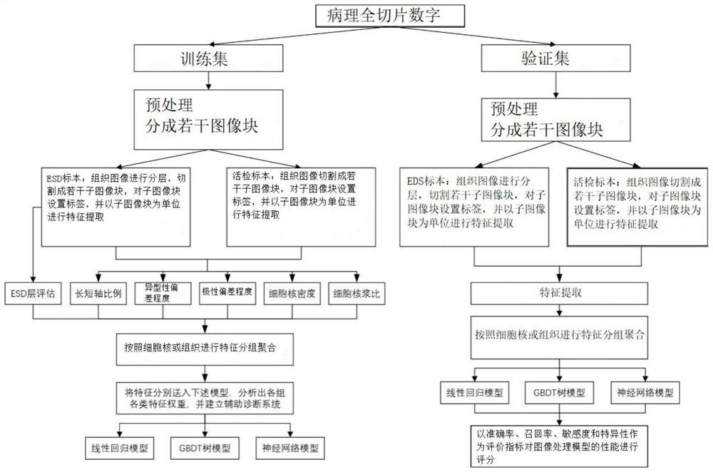
[0023] This embodiment discloses a method for processing an image of esophageal squamous epithelial dysplasia, which includes the following steps:

[0024] S1 divides pathological full-section digital images into a training set and a verification set, uses the data in the training set to train the model, uses the data in the verification set to select the model, and preprocesses the images in the training set and the verification set respectively.

[0025] The preprocessing method is to perform data enhancement on all pathological full-section digital images, and make binary masks for the effective tissue area and the labeled lesion tissue area respectively. The data enhancement methods for the training set include color gamut migration, rotation, and cropping. The set of data augmentation methods includes gamut shift. Among them, the pathological full-section digital image used in this embodiment does not need to be manually accurately labeled, but only briefly outlines the l...

Embodiment 2

[0049] Based on the same inventive concept, this embodiment discloses a processing system for images of esophageal squamous epithelial dysplasia, including:

[0050] The preprocessing module is used to divide the digital image of the pathological full slice into a training set and a verification set, and preprocess the images in the training set and the verification set respectively;

[0051] A feature extraction module for dividing all preprocessed images into ESD specimen images and living specimen images, performing histological classification on ESD specimens, stratifying ESD specimens, evaluating EDS layers, and extracting ESD specimen images and Eigenvalues ​​of biopsy specimen images;

[0052] The model training module is used to group and aggregate the feature values ​​of the ESD sample images and biopsy sample images of the images in the training set according to the nucleus or tissue feature grouping, and use the feature values ​​after feature grouping and aggregatio...

Embodiment 3

[0056] Based on the same inventive concept, this embodiment discloses a computer-readable storage medium, on which a computer program is stored, and the computer program is executed by a processor to realize any of the above-mentioned esophageal squamous epithelial dysplasia images processing method.

[0057] Those skilled in the art should understand that the embodiments of the present application may be provided as methods, systems, or computer program products. Accordingly, the present application may take the form of an entirely hardware embodiment, an entirely software embodiment, or an embodiment combining software and hardware aspects. Furthermore, the present application may take the form of a computer program product embodied on one or more computer-usable storage media (including but not limited to disk storage, CD-ROM, optical storage, etc.) having computer-usable program code embodied therein.


PUM


Abstract

The invention relates to an esophageal squamous epithelium dysplasia image processing method and system and a medium, and the method comprises the steps: dividing a pathological full-slice digital image into a training set and a verification set, and carrying out the preprocessing of the training set and the verification set; dividing the preprocessed image into an ESD specimen image and a living specimen image, layering an ESD specimen, evaluating an EDS layer, and extracting feature values of the ESD specimen image and the biopsy specimen image; carrying out feature grouping and aggregation on the feature values of the images in the training set according to cell nucleuses or tissues, and training an image processing model by adopting the feature values subjected to feature grouping and aggregation to obtain an optimal image processing model; verifying the optimal image processing model by using the feature values of the images in the verification set; and inputting the to-be-detected image into the optimal image processing model to obtain the type of the atypical hyperplasia cells or tissues in the to-be-detected image. The method solves the problem of graphic feature extraction of cell tissues in the absence of precisely marked medical images.

Description

technical field [0001] The invention relates to a processing method, system and medium for images of esophageal squamous epithelial dysplasia, and belongs to the technical field of medical image processing. Background technique [0002] Esophageal cancer is one of the most common malignant tumors in the world, and my country is one of the countries with the highest incidence of esophageal cancer. Most esophageal cancers are diagnosed at an advanced stage, resulting in a poor overall prognosis. If early detection, early diagnosis, and early treatment are available, the 5-year survival rate of patients can exceed 90%. Due to different degrees of differentiation, the cell morphology and tissue structure of tumors are different from the corresponding normal tissues to varying degrees, and this difference is called atypia. Esophageal squamous epithelial dysplasia is a kind of precancerous lesion, which is atypical cells in the esophageal squamous epithelium without invasion. Es...

Claims


Application Information

Patent Timeline
Patent Type & Authority Applications(China)
IPC IPC(8): G06K9/62G06K9/46G16H30/20G16H50/20
CPCG16H50/20G16H30/20G06V10/40G06F18/241G06F18/214Y02A90/10
Inventor 金木兰王莹祝闯石中月刘军段佳佳罗毅豪
Owner BEIJING CHAOYANG HOSPITAL CAPITAL MEDICAL UNIV
Why Eureka
Eureka Blog
Learn More