CT image segmentation method based on improved AU-Net network

A CT image and network technology, applied in the field of image processing, can solve problems such as uneven distribution of target areas, unfavorable extraction of image features, and impact on segmentation effects.

Active Publication Date: 2021-06-08
CHONGQING UNIV OF POSTS & TELECOMM
View PDF4 Cites 8 Cited by
  • Summary
  • Abstract
  • Description
  • Claims
  • Application Information

AI Technical Summary

Problems solved by technology

[0005] There are limitations when the above-mentioned prior art is directly applied to CT image segmentation of cerebral hemorrhage; due to the complexity of the structure of the human brain, the pixel features of the cerebral hemorrhage area on the CT image are very similar to the pixel features of the skull. , the pixels in the skull will interfere with the lesion area; cerebral h

Method used


Image

  • CT image segmentation method based on improved AU-Net network
  • CT image segmentation method based on improved AU-Net network
  • CT image segmentation method based on improved AU-Net network

Examples


Embodiment Construction

[0047] The following will clearly and completely describe the technical solutions in the embodiments of the present invention with reference to the accompanying drawings in the embodiments of the present invention. Obviously, the described embodiments are only some, not all, embodiments of the present invention. Based on the embodiments of the present invention, all other embodiments obtained by persons of ordinary skill in the art without making creative efforts belong to the protection scope of the present invention.

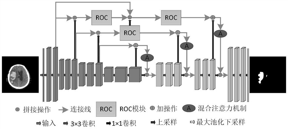
[0048] A CT image segmentation method based on the improved AU-Net network, such as Figure 7 As shown, the method includes: obtaining the brain CT image to be segmented, and preprocessing the obtained brain CT image; inputting the processed image into the trained improved hybrid attention mechanism network AU-Net for image recognition and segmentation, The segmented CT image is obtained; the brain hemorrhage area is identified according to the segmented brain...


PUM


Abstract

The invention belongs to the field of image processing, and particularly relates to a CT image segmentation method based on an improved AU-Net network, and the method comprises the steps: obtaining a to-be-segmented brain CT image, and carrying out the preprocessing of the obtained brain CT image; inputting the processed image into the trained improved AU-Net network for image recognition and segmentation to obtain a segmented CT image; identifying a cerebral hemorrhage area according to the segmented brain CT image. The improved AU-Net network comprises an encoder, a decoder and a hopping connection part. Aiming at the problem of low segmentation precision caused by large size and shape difference of the hemorrhage part of the cerebral hemorrhage CT image, the invention provides a coding-decoding-based structure, and a residual octave convolution module is designed in the structure, so that the model can segment and identify the image more accurately.

Description

technical field [0001] The invention belongs to the field of image processing, in particular to a CT image segmentation method based on an improved AU-Net network. Background technique [0002] Cerebral hemorrhage refers to primary cerebral parenchymal hemorrhage, also known as cerebral hematoma, which can be detected by magnetic resonance imaging (Magnetic Resonance Imaging, MRI), computed tomography (Computed-Tomography, CT), ultrasound (Ultrasound, US) and other medical imaging techniques The obtained organ anatomy map can objectively reflect the pathological changes of the patients. Clinically, cerebral hemorrhage appears as a bright area on CT images, and normal brain soft tissues appear as black on CT images. The doctor judged the hemorrhage volume and other relevant indicators based on the cerebral hemorrhage area shown on each CT image combined with experience. Use image segmentation technology to segment the target area of ​​each cerebral hemorrhage CT image, calc...

Claims


Application Information

Patent Timeline
IPC IPC(8): G06T7/11G06N3/04G06N3/08G06T5/30G06T5/50
CPCG06T7/11G06T5/30G06T5/50G06N3/084G06T2207/10081G06T2207/20081G06T2207/20221G06T2207/30016G06N3/045
Inventor 胡敏周秀东黄宏程
Owner CHONGQING UNIV OF POSTS & TELECOMM
Why Eureka
Try Eureka