

Image processing method, device and equipment and readable storage medium
An image processing and initial image technology, applied in the field of medical image processing, can solve the problems of high scanning times, low efficiency, lack of efficient and fast automatic bone removal methods, etc., and achieve the effect of reducing noise data
- Summary
- Abstract
- Description
- Claims
- Application Information
AI Technical Summary
Problems solved by technology
Method used
Image

Examples
Embodiment 1
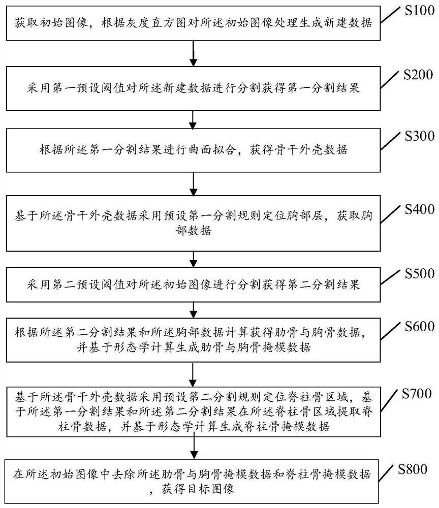
[0066] Embodiment 1: This embodiment discloses an image processing method, which is applied to a computerized tomography blood vessel analysis system for realizing bone removal in cardiovascular angiography images. Refer to figure 1 , specifically including the following steps:
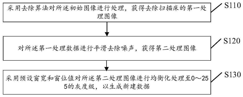
[0067] S100: Acquire an initial image, and generate new data by processing the initial image according to the grayscale histogram;
[0068] In this embodiment, the grayscale histogram reflects the relationship between the frequency of occurrence of each grayscale pixel in an image and the grayscale level, with the grayscale as the abscissa and the frequency as the ordinate, and the drawing frequency is the same as the grayscale The relationship image is the grayscale histogram of the image. According to the grayscale histogram, the grayscale distribution of the image can be reflected, and the threshold of image binarization can also be determined at the same time, so as to determine the binary image o...
Embodiment 2
[0110] Embodiment 2: This embodiment provides an image processing device 8, refer to Figure 7 , including the following:
[0111] A preprocessing module 81, configured to acquire an initial image, and generate new data by preprocessing the initial image according to the grayscale histogram;
[0112] The first segmentation module 82 is configured to segment the newly created data by using a first preset threshold to obtain a first segmentation result;
[0113] A backbone shell data generating module 83, configured to obtain backbone shell data by fitting according to the first segmentation result;
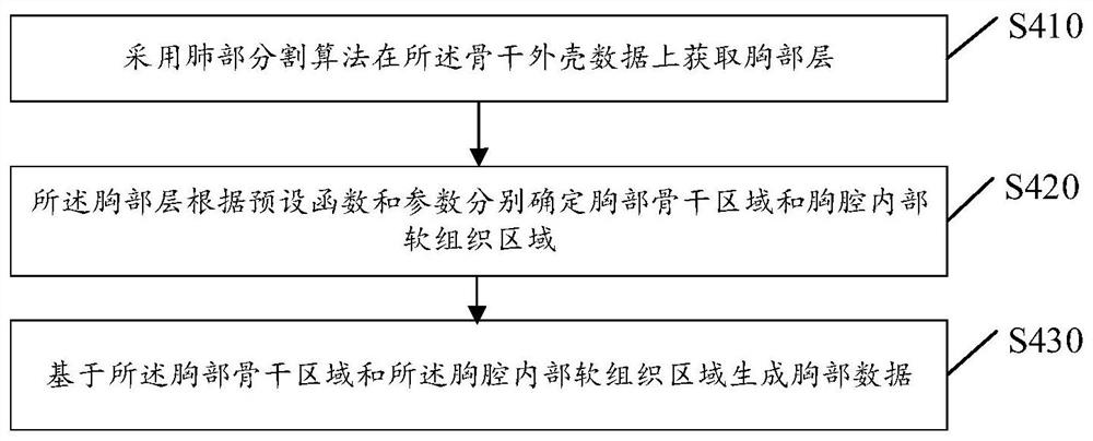
[0114] The chest data generation module 84 is configured to locate the chest layer by using a preset first segmentation rule based on the backbone shell data, and obtain chest data; specifically, the first segmentation rule includes lung segmentation and a preset function.
[0115] The second segmentation module 85 is configured to segment the initial image using a second preset ...
Embodiment 3
[0120] Embodiment 3: In order to achieve the above purpose, the present invention also provides a computer device 9, see Figure 8 , the computer equipment may include a plurality of computer equipment, and the components of the GPU-based image processing device 8 of Embodiment 2 may be dispersed in different computer equipment 9, and the computer equipment 9 may be a smart phone, a tablet computer, a notebook for executing a program Computers, desktop computers, rack servers, blade servers, tower servers or rack servers (including independent servers, or server clusters composed of multiple servers), etc. The computer equipment in this embodiment at least includes but is not limited to: a memory 91, a processor 92, and an image processing device 8 for caching GPUs that can be connected to each other through a system bus, such as Figure 8 shown. It should be pointed out that, Figure 8 Only a computer device is shown with the components - but it should be understood that im...
PUM
 Abstract
Description
Claims
Application Information