Mucosal melanoma cell and application thereof
A technology for melanoma cells and melanoma, applied in the field of mucosal melanoma cells, can solve the problems of difficult to obtain samples, cell aging, growth stagnation, etc.
- Summary
- Abstract
- Description
- Claims
- Application Information
AI Technical Summary
Problems solved by technology
Method used
Image

Examples
Embodiment 1
[0070] Example 1 Establishment of cell line
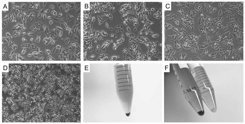
[0071] The sources of specimens were the oral and maxillofacial head and neck of the Ninth People's Hospital of Shanghai Jiaotong University, an example of a 67-year-old female mucosal melanoma, primary stove is located in the lower teeth mucosa. Before the freezing activity, the patient and the guardian agreed to sign the informed consent, and the postoperative pathological diagnosis was: mucosa melanoma. Organizational dyeing is shown in Figure (9A).
[0072] Specific culture method:
[0073] The biopsy sample quickly loaded into the centrifuge tube containing the commercial tissue storage liquid (MACS, Germany). The centrifuge tube is loaded into the ice cartridge to the laboratory.
[0074] In the biosafety cabinet, the tissue was transferred to the culture dish, and the PBS immersion tissue containing amphotericin B (500 ng / ml) and 5% pyroxin double anti-anti-double anti-antigen was about 10 minutes. The mucosal tissue was remov...
Embodiment 2
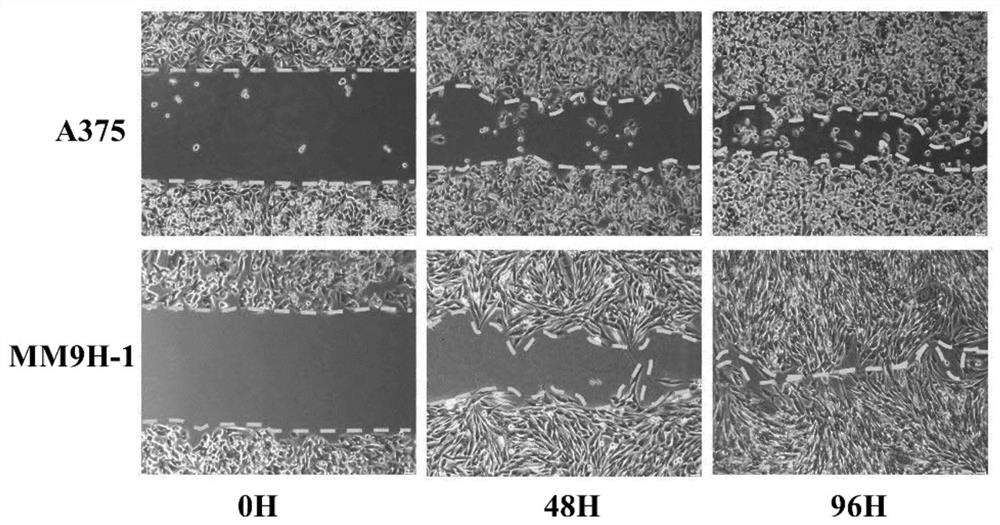
[0080] Example 2 Cell scratches experiment
[0081] Cell scratches showed that MM9H-1 migration capacity is stronger than classic human skin melanoma cells A375 ( image 3 ).
[0082] Cell scratches experiments use wound healing plugs (IBIDI, Germany, # 81176) experiment, which can generate an neat scale area. The specific steps are as follows: Put the plug in 12-well plates, and the MM9H-1 cell digestion is prepared as a suspension of about 5 x 10⁶ / ml cell suspension, inoculated in the plug-in, 80 microliter cytosin suspensions per well; 24 After the hour, under the mirror, the cells were observed, about 90 ~ 100% aggregation, remove the plug, add 500 microlitrated serum culture based on 12-well plate; microscope, as 0 baseline; observe once every 24 hours, take pictures, Determination of cell migration distance between scratches.
Embodiment 3
[0083] Example 3 Cell line melanoma-related marker staining
[0084] MM9H-1 of human mucosa tumor cells MM9H-1 was identified by immunofluorescence technology MM9H-1, MMB45, MELAN-A, S100A6, S100B for identification. Such as Figure 4 As shown, human mucosa melanoma cells MM9H-1 expression marker HMB45, MELAN-A, S100A6. MM9H-1 was detected by MM9H-1 with melanoma hybrid label antibody in P50 generation, HMB45, Melan-A and other markers were stable ( Figure 5 ).
[0085]MM9H-1 cells were inoculated into a cop-focus peani having a diameter of 12 mm. After 24 h, the culture solution, PBS was washed twice, and the pre-cooling 4% polymethyledehyde was fixed for 30 min, and 5 min × 3 times at room temperature PBS. 0.1% Triton X-100 treatment 10min, room temperature PBS wash 5 min × 3 times, 5% goat blood cleaning closed 30 min, add dilution imperative (HMB45, Melan-a, S100A6) placed in a wet cartridge, 4 ° C overnight, room temperature PBS wash To the anti-anti-affordable, 10 min × 3 tim...
PUM
 Abstract
Description
Claims
Application Information
 - R&D
- Intellectual Property
- Life Sciences
- Materials
- Tech Scout
- Unparalleled Data Quality
- Higher Quality Content
- 60% Fewer Hallucinations
Browse by: Latest US Patents, China's latest patents, Technical Efficacy Thesaurus, Application Domain, Technology Topic, Popular Technical Reports.
© 2025 PatSnap. All rights reserved.Legal|Privacy policy|Modern Slavery Act Transparency Statement|Sitemap|About US| Contact US: help@patsnap.com