Microvascular distortion degree quantification method for gastric mucosa dyeing amplification imaging

A technology of distortion degree and quantification method, applied in the field of image processing in the medical field, can solve the problems of inability to automate and batch, and achieve the effect of improving agility, reliability and accuracy.

Active Publication Date: 2021-08-03
WUHAN UNIV
View PDF5 Cites 7 Cited by
  • Summary
  • Abstract
  • Description
  • Claims
  • Application Information

AI Technical Summary

Problems solved by technology

However, the quantification process of the degree of vascular distortion in the paper involves a variety of software, such as using AWvolume share4.0 works

Method used


Image

  • Microvascular distortion degree quantification method for gastric mucosa dyeing amplification imaging
  • Microvascular distortion degree quantification method for gastric mucosa dyeing amplification imaging
  • Microvascular distortion degree quantification method for gastric mucosa dyeing amplification imaging

Examples


Embodiment Construction

[0042] The present invention will be described in further detail below in conjunction with the accompanying drawings and specific embodiments, and the implementation scope of the present invention is not limited thereto.

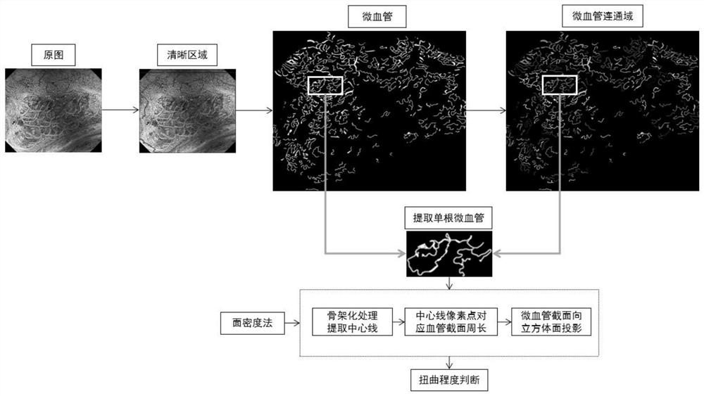
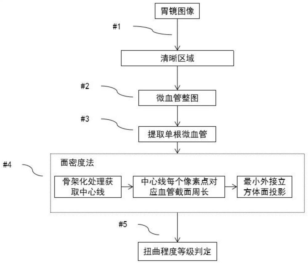
[0043] Please refer to figure 1 , a schematic diagram of the implementation of the method for quantifying the degree of microvascular distortion in mucosal staining magnification imaging of the present invention. Such as figure 1 As shown, a method for quantifying the distortion degree of microvessels in gastric mucosa staining and magnification imaging of the present invention includes the surface density method.

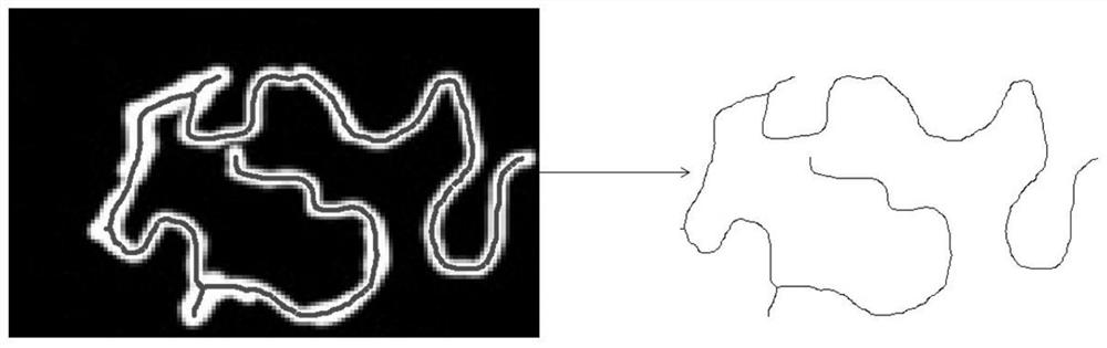
[0044] Please refer to figure 2 , which takes an embodiment of the method for quantifying the degree of microvessel distortion of gastric mucosa staining and magnification imaging as an example to illustrate the establishment of the process flow of the method for quantifying the degree of distortion of gastric microvessels in the present inven...


PUM


Abstract

The invention discloses a microvascular distortion degree quantification method for gastric mucosa dyeing amplification imaging. The method comprises an image segmentation method, a blood vessel center line extraction method and a surface density method. The image segmentation method is used for extracting a clear area and a microvessel whole image in the gastroscope image; the blood vessel center line extraction method is used for extracting the center line of each microvessel and calculating the perimeter of the microvessel corresponding to a pixel point on the center line; according to the surface density method, calculating the surface density by projecting the perimeter of the microvessel to the minimum circumscribed cubic surface of the microvessel; and finally, weighting the surface densities of different surfaces of the minimum microvessel circumscribed cube to obtain a microvessel distortion degree coefficient, and then judging the microvessel distortion degree grade according to the distortion degree coefficient.

Description

technical field [0001] The invention relates to the technical field of image processing in the medical field, in particular to a method for quantifying the twisting degree of microvessels in gastric mucosa staining and magnification imaging. Background technique [0002] Gastric cancer is the fifth most common cancer in the world, and it is the third leading cause of cancer death. The five-year survival rate of advanced gastric cancer is only 30%, while the five-year survival rate of early gastric cancer is as high as 90%. The advent of chromoendoscopy established the endoscopic diagnosis method for early gastric cancer. The combined application of magnifying gastroscopy and electronic chromoendoscopy can observe the tiny blood vessel structure and the fine structure of mucosal surface that cannot be observed by ordinary gastroscopy, which provides conditions for endoscopic diagnosis of early gastric cancer. Vascular dysmorphia is an important condition for the diagnosis o...

Claims


Application Information

Patent Timeline
IPC IPC(8): G06T7/00G06T7/11G06T7/187G06T7/62G06T3/40G06T5/00
CPCG06T7/0012G06T7/62G06T7/11G06T7/187G06T5/002G06T3/40G06T2207/10068G06T2207/30092G06T2207/30101
Inventor 于红刚吴练练胡珊李昊朱益洁徐铭陶逍张梦娇李佳董泽华张晨霞杜泓柳陈弟何鑫琦黄丽商任铎
Owner WUHAN UNIV
Why Eureka
Try Eureka