Preparation method of ultra-small bionic nanoparticles based on erythrocyte membrane
A red blood cell membrane, nanoparticle technology, applied in nanotechnology, nanotechnology, nanomedicine, etc., can solve the problems of product residue, low efficiency, difficult to obtain, etc., and achieve the effect of stable reaction and mild environment.
- Summary
- Abstract
- Description
- Claims
- Application Information
AI Technical Summary
Problems solved by technology
Method used
Image

Examples
Embodiment 1
[0044] Preparation and characterization of PLGA particles
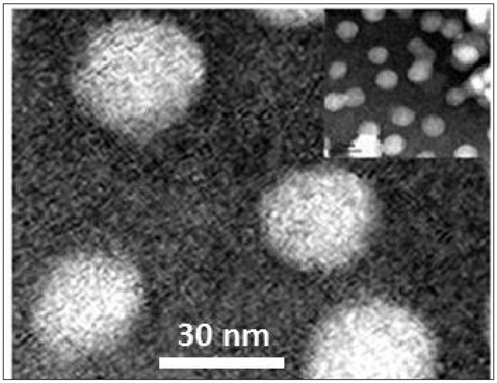
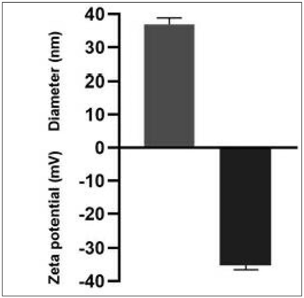
[0045] Prepare PLGA particles by nano-co-precipitation method: Dissolve PLGA (0.67 dL / g) with a viscosity of 50:50 and carboxy-terminal modification in acetone to 7 mg / mL for use. The above 1 mL PLGA acetone solution was quickly added to 3 mL PH=8.0 Tris-HCl deionized water pre-cooled at 4°C, and then the stirring speed was kept at 600 rpm / min until the acetone was completely evaporated. The collected PLGA nanoparticles were prepared and collected for basic physical characterization, including observing the particle morphology with a transmission electron microscope and detecting the hydraulic size and surface potential of the particles with a particle size potentiometer. figure 1 The TEM results show that the size of PLGA nanoparticles is about 30 nanometers; figure 2 The particle size potentiometer test results show that the hydraulic size of PLGA nanoparticles is about 35 nanometers, and the surface potential is ...
Embodiment 2
[0047] Extraction and purification of ICR mouse erythrocyte membrane
[0048] Erythrocyte extraction from ICR mice: Blood was drawn through the cheek and mixed with 10 mM EDTA in PBS at a volume ratio of 1:1. Centrifuge at 3500rpm for 15min, carefully remove the serum and white blood cell layer, and take the red blood cell layer. Red blood cells were added to deionized water at a volume ratio of 1:6, mixed well and incubated in an ice bath for 30 min. Subsequently, a certain volume of 20XPBS was added to adjust the erythrocyte membrane solution to 1X PBS, and then centrifuged at 13800rpm for 10min to remove hemoglobin in the supernatant. The above process was repeated 3 to 4 times until no hemoglobin was detected in the centrifuged supernatant. The supernatant was removed, the red blood cell membrane was resuspended with deionized water, and the membrane protein concentration was measured by BCA method, and finally stored at -80°C. image 3 Represents red blood cells before...
Embodiment 3
[0050] Preparation, characterization and in vitro stability determination of erythrocyte membrane-encapsulated biomimetic nanoparticles (PLGA@RBC)
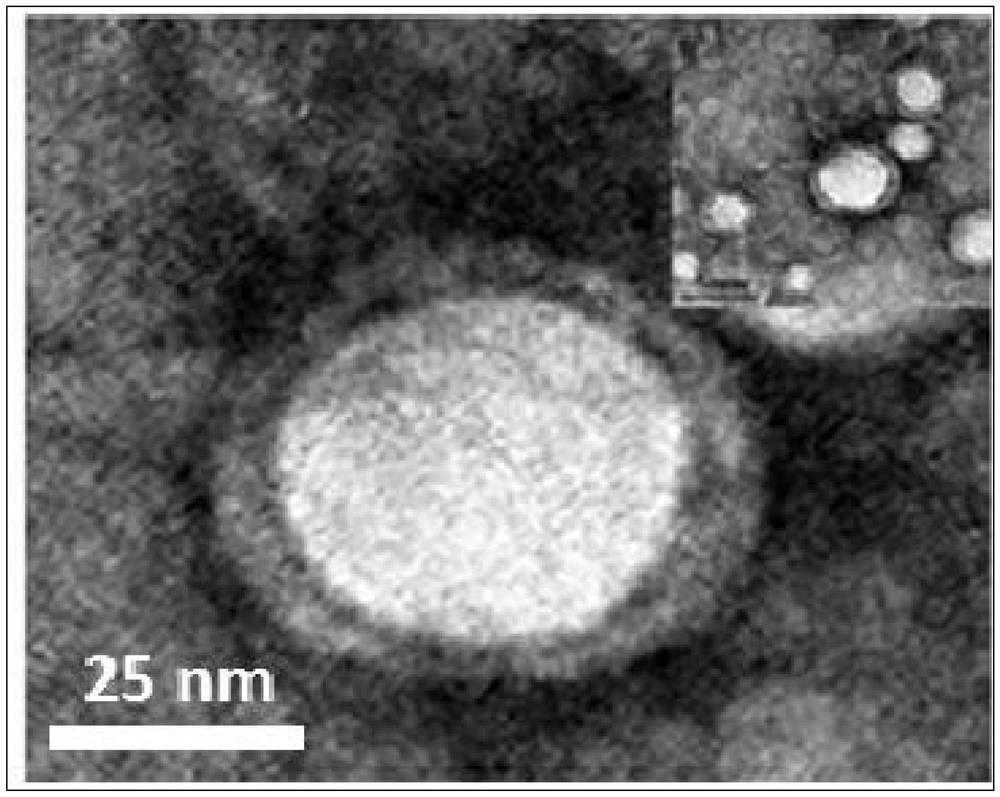
[0051]Dilute PLGA:RBC to 1.5mL according to 1:2 (w / w), then add it into a 20mL glass bottle, and ultrasonicate in a water bath for 5min to prepare biomimetic nanoparticles wrapped in red blood cell membrane, which is designated as PLGA@RBC. The prepared and collected PLGA@RBC were subjected to basic physical characterization, including the observation of particle morphology by transmission electron microscopy and the detection of the hydraulic size and surface potential of the particles by a particle size potentiometer. image 3 The TEM results show that PLGA@RBC presents a core-shell structure with a size of about 50 nanometers; Figure 4 The particle size potentiometer test results show that the hydraulic size of PLGA@RBC is about 60 nanometers, and the surface potential is about -27mV. Figure 5 It was shown that PLGA@RBC has ...
PUM
| Property | Measurement | Unit |
|---|---|---|
| surface potential | aaaaa | aaaaa |
| surface potential | aaaaa | aaaaa |
Abstract
Description
Claims
Application Information
 - R&D Engineer
- R&D Manager
- IP Professional
- Industry Leading Data Capabilities
- Powerful AI technology
- Patent DNA Extraction
Browse by: Latest US Patents, China's latest patents, Technical Efficacy Thesaurus, Application Domain, Technology Topic.
© 2024 PatSnap. All rights reserved.Legal|Privacy policy|Modern Slavery Act Transparency Statement|Sitemap