Eureka AIR delivers breakthrough ideas for toughest innovation challenges, trusted by R&D personnel around the world.

Pressing hemostasis device for nursing after angiocardiography

A cardiovascular and installation groove technology, which is applied in the field of pressing hemostatic devices for nursing after cardiovascular angiography, can solve the problems of reducing the work efficiency of medical staff, reducing the flexibility of hemostatic devices, and the hemostatic plate cannot be fully fitted, so as to increase convenience , increase practicability, and solve a single non-adjustable effect

Pending Publication Date: 2021-10-08
PEOPLES HOSPITAL OF XINJIANG UYGUR AUTONOMOUS REGION
View PDF9 Cites 0 Cited by
  • Summary
  • Abstract
  • Description
  • Claims
  • Application Information

AI Technical Summary

Problems solved by technology

[0004] The purpose of the present invention is to provide a compression hemostasis device for nursing after cardiovascular angiography, to solve the problem that the current compression hemostasis device proposed in the background technology is mostly fixed by screwing the bolt when it is in use. The fixed method takes more working time for medical staff, which not only reduces the work efficiency of medical staff, but also increases the cumbersomeness of the use of the hemostatic device; most of the current pressing hemostatic devices are not convenient for medical staff to stop bleeding at different positions on the patient's surface The use of the hemostatic device reduces the flexibility of the use of the hemostatic device. At the same time, some bleeding positions require medical staff to manually press to stop the bleeding, which increases personnel consumption and reduces the practicability of the hemostatic device. Most of the hemostatic plates used in the existing hemostatic device The arc structure is fixed, because the arc generated at the position of the patient's body surface does not match the arc of the hemostatic plate used, which will cause the hemostatic plate to not fully fit the patient's surface, reducing the hemostatic effect and reducing the patient's hemostatic effect. Questions about comfort of use

Method used


Image

  • Pressing hemostasis device for nursing after angiocardiography
  • Pressing hemostasis device for nursing after angiocardiography
  • Pressing hemostasis device for nursing after angiocardiography

Examples


Embodiment 1

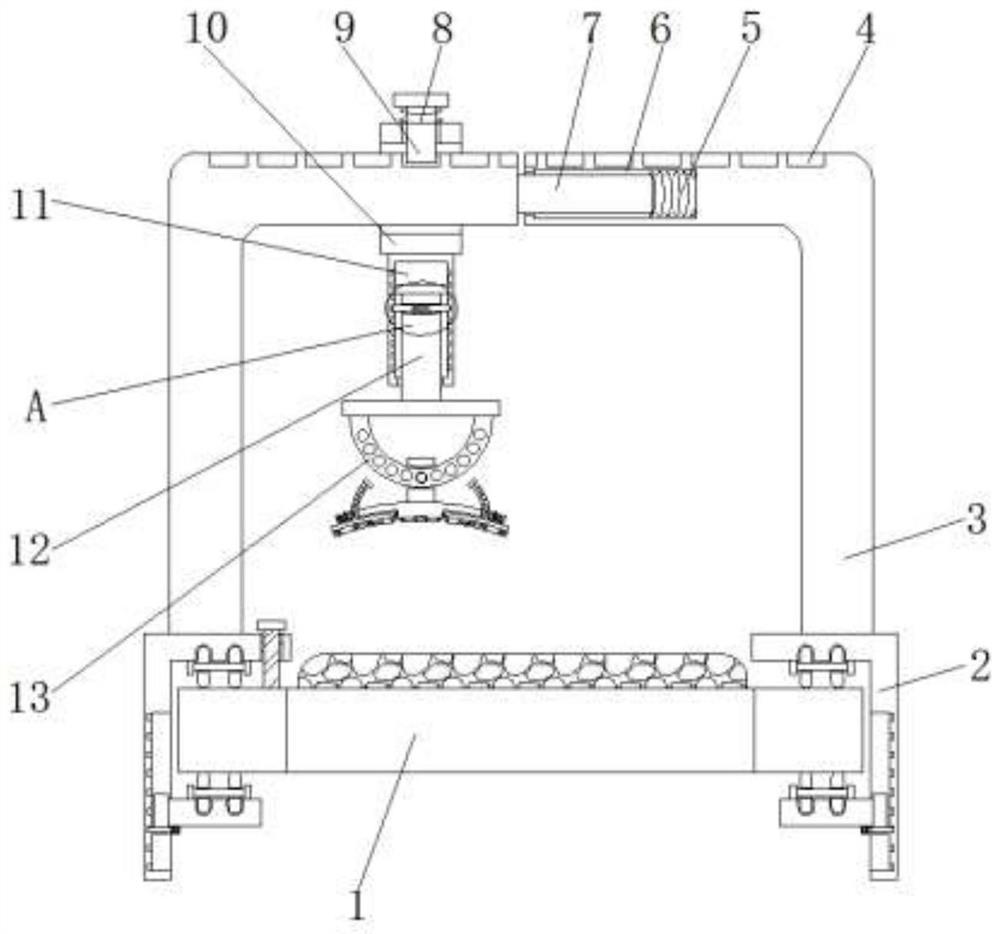
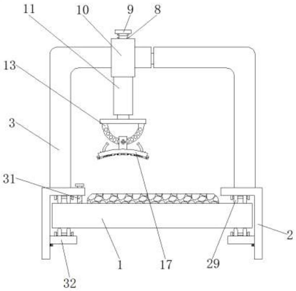
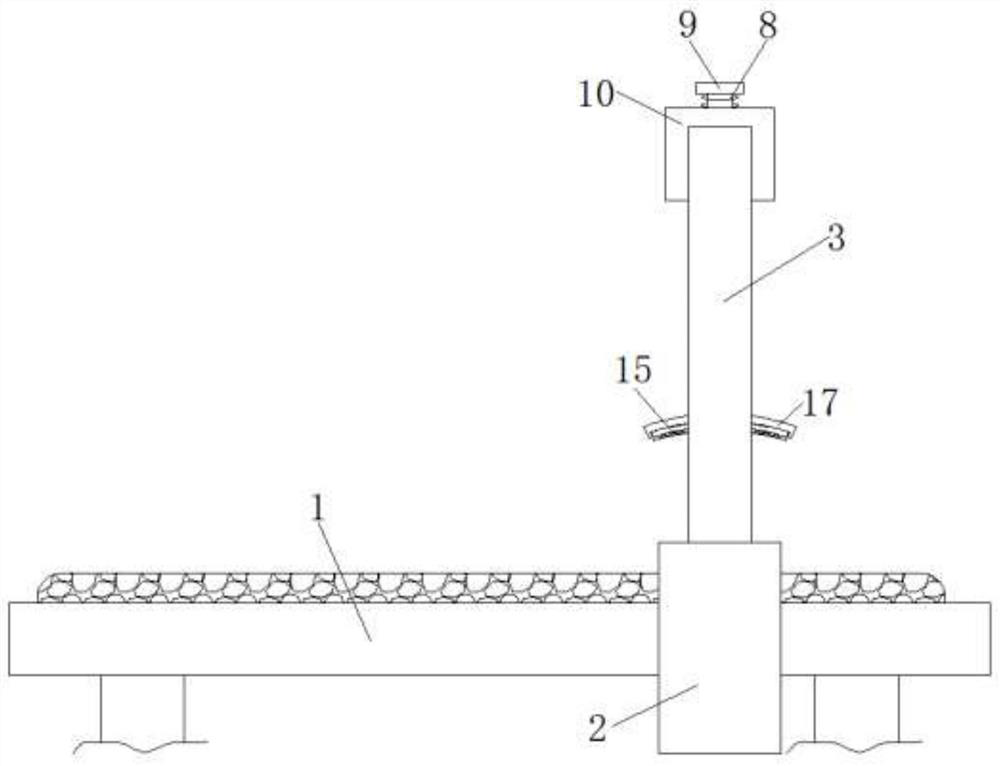
[0045] Example 1, such as Figure 5 , 8 As shown, when installing, you can pull the L-shaped connecting rod 3 and the above parts first, so that it drives the connecting rod 7 to move inside the first installation groove 6 to stretch the first spring 5. After the pulling is completed, Place the L-shaped connecting rod 3 and its above parts on both sides of the operating bed 1, and then drive the L-shaped connecting rod 3 and its components to recover through the elastic restoring force of the first spring 5. After the placement is completed, pull the fixing rod 36 to make it move inside the slider 34 to stretch the fifth spring 35, so that the fixing rod 36 can be moved out to the inside of the fixing groove 33 to release the fixing of the mounting plate 32, and then pull the mounting plate 32 to drive it to slide The block 34 slides to the designated position inside the chute 28, and then loosens the fixed rod 36 through the elastic restoring force of the fifth spring 35, wh...

Embodiment 2

[0046] Example 2, such as figure 1 , 6 As shown, when it is necessary to stop bleeding at the bleeding position of the patient, the slide bar 12 and its parts above can be directly pulled to slide down inside the sleeve 11, and at the same time, the movement of the slide bar 12 and its parts above can make the The sleeve 11 pushes the limit rod 27 to move inside the third installation groove 25 to compress the fourth spring 26, so that the limit rod 27 can be disengaged from the inside of the limit groove 24 to release the sliding rod 12 and its above components. In order to reduce the wear and tear of personnel and improve the practicability of the device, the sliding bar 12 pushes the arc-shaped hemostatic plate 17 and the above parts to press the patient's bleeding site to stop bleeding.

[0047] Working principle: First, according to the width of the operating bed 1, the L-shaped connecting rod 3 and its above parts can be pulled first, so that it can drive the connecting...


PUM


Abstract

The invention discloses a pressing hemostasis device for nursing after angiocardiography. The pressing hemostasis device comprises an operating bed; L-shaped clamping plates are arranged on the two sides of the operating bed; clamping assemblies are arranged on the sides, close to each other, of the two L-shaped clamping plates; L-shaped connecting rods are arranged at the tops of the two L-shaped clamping plates; the lower portions of the sides, close to each other, of the two L-shaped connecting rods are provided with connecting rods and first mounting grooves correspondingly; and the connecting rods extend into the first mounting grooves. According to the pressing hemostasis device for nursing after the angiocardiography, through the mutual cooperation of the operating bed, the L-shaped connecting rods, the connecting rods, the first mounting grooves, a first spring, a fixing rod, a sliding block, a fifth spring, a fixing groove, a mounting plate, a sliding groove and the L-shaped clamping plates, medical staff can conveniently and quickly mount the hemostasis device through cooperation of the parts, and the medical staff does not need to screw and fix the hemostasis device through a bolt. Therefore, the working steps of the medical staff are reduced, the working efficiency of the medical staff is improved, and the use convenience of the hemostasis device is improved.

Description

technical field [0001] The invention relates to the technical field of compression hemostasis devices, in particular to a compression hemostasis device for nursing after cardiovascular angiography. Background technique [0002] Traditional angiography, a technique in which the blood vessels are visualized by injecting a contrast agent, is performed in a cardiac catheterization in order to identify any heart chambers, great vessels, or coronary vessels. After performing the percutaneous puncture operation on angiography, the puncture site needs to be hemostasis. At this time, manual compression or an auxiliary hemostatic device are generally used for hemostasis. [0003] When the current pressing hemostatic device is installed, most of it is fixed by screwing the bolt. This kind of fixing method takes more working time for the medical staff, which not only reduces the work efficiency of the medical staff, but also increases the hemostasis. The use of the device is cumbersome...

Claims


Application Information

Patent Timeline
IPC IPC(8): A61B17/12
CPCA61B17/12A61B2017/12004
Inventor 高燕宁娜陈义董丹萍热衣拉·买买提柯晓琴熊英谢芳
Owner PEOPLES HOSPITAL OF XINJIANG UYGUR AUTONOMOUS REGION
Why Eureka
Eureka Blog
Learn More