
Animal belly window manufacturing method suitable for optical in-vivo organ imaging

A manufacturing method and imaging technology, applied in the field of biomedical imaging, can solve the problems of inability to simulate a closed abdominal cavity, high risk of animal infection and death, etc.

Pending Publication Date: 2021-10-19
NANJING RAYGEN HEALTH CO LTD
View PDF0 Cites 0 Cited by
  • Summary
  • Abstract
  • Description
  • Claims
  • Application Information

AI Technical Summary

Problems solved by technology

[0003] Aiming at the above defects of the prior art, the present invention proposes a method for making an animal ventral window suitable for optical imaging of living animal organs. Using this method, one surgical operation can be performed, and then multiple, continuous long-term imaging can be performed to solve the problem. In the past, only one imaging experiment could be done for one operation, which could not simulate the closed abdominal cavity state, and could only present the microscopic morphology of the exposed organs, and the risk of postoperative infection and death of animals was higher.

Method used


Image

  • Animal belly window manufacturing method suitable for optical in-vivo organ imaging
  • Animal belly window manufacturing method suitable for optical in-vivo organ imaging
  • Animal belly window manufacturing method suitable for optical in-vivo organ imaging

Examples


Embodiment Construction

[0009] In this specific embodiment, the actin-GFP transgenic mouse is taken as an example of an experimental animal, and the specific steps are as follows:

[0010] 1. Preparation before experiment

[0011] (1), experimental animal anesthesia

[0012] The mice prepared for the operation experiment were placed in the induction anesthesia cabin of the anesthesia machine, the anesthesia index was adjusted to 3%-5%, and the induction anesthesia was carried out, and the process lasted about two minutes. The appropriate performance for judging the depth of anesthesia in mice is that the mice breathe evenly, and the frequency is every second. Move the mouse from the anesthesia cabin to the continuous anesthesia mask of the anesthesia machine, connect the mouse incisors to the small holes of the mask and tightly cover the mouth and nose of the mouse, and adjust the anesthesia index to 0.3%-0.5%. The state of the mouse is fine-tuned in real time to ensure that the breathing rate of t...


PUM


Abstract

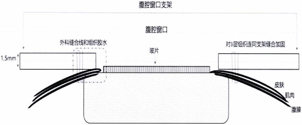
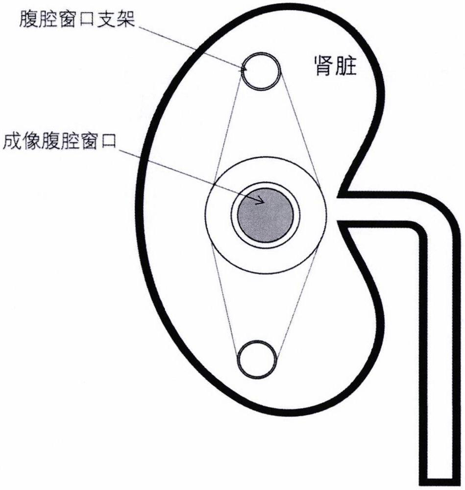
The invention discloses an animal belly window manufacturing method suitable for optical in-vivo organ imaging. The method comprises the following steps: (1) covering the surface of an organ to be imaged with a circular slide, sealing the periphery of the circular slide with tissue glue, and adhering the circular slide to the surface of the organ to be imaged, so as to make the circular slide and the organ to be imaged tightly attached; (2) bonding the imaging organ, peritoneum, muscle and skin which are not covered by the round slide by using tissue glue, and bonding the outside of the skin with a window fixing plate; and (3) finally, suturing and reinforcing the skin, muscle and peritoneum three-layer tissues and the window fixing plate by using surgical sutures. The imaging window is firmly installed on the surface of the imaging organ, and the requirement of living animal in-vivo organ imaging is met.

Description

technical field [0001] The invention relates to a method for making an animal ventral window suitable for optical in vivo organ imaging, and belongs to the field of biomedical imaging. Background technique [0002] At present, in the field of subcellular resolution imaging of biological samples, specimen fixation and microscopic imaging after pathological sections are mostly used; or necessary operations such as anesthetizing experimental animals, fixing the body, and fully exposing the imaging organs are suitable for one-time imaging. Sexual imaging experiments, and the animal mortality rate is high, it is impossible to carry out long-term, multiple tracking experiments. Contents of the invention [0003] Aiming at the above defects of the prior art, the present invention proposes a method for making an animal ventral window suitable for optical imaging of living animal organs. Using this method, one surgical operation can be performed, and then multiple, continuous long-...

Claims


Application Information

Patent Timeline
Patent Type & Authority Applications(China)
IPC IPC(8): A61D1/00A61B5/00
CPCA61D1/00A61B5/0071A61B5/0077
Inventor 杨光亮赵婷刘辉
Owner NANJING RAYGEN HEALTH CO LTD
Learn More