

U-Net fundus retinal blood vessel image segmentation method and device based on improvement
A retinal blood vessel and image segmentation technology, applied in image analysis, image data processing, neural learning methods, etc., can solve the problems of low contrast information of retinal blood vessels in the fundus, low precision of fine-grained blood vessel areas, etc., and achieve low accuracy, Good image-related features, the effect of improving robustness
Active Publication Date: 2021-11-02
BEIJING UNION UNIVERSITY
View PDF11 Cites 1 Cited by
- Summary
- Abstract
- Description
- Claims
- Application Information
AI Technical Summary
Problems solved by technology
[0005]
However, the accuracy of the existing methods for the segmentation of fine-grained blood vessel regions is still relatively low, especially for the low-contrast information of retinal blood vessels in the fundus, which is difficult to identify. How to better locate and segment microvessels is a problem that needs to be solved at present.
Method used
View moreImage
Smart ImageViewing Examples

Examples
specific Embodiment
[0043] Figure 1 to Figure 6 A specific embodiment of a method for segmenting retinal blood vessels based on an improved U-Net fundus image of the present invention is shown, including the following steps:
[0044] Step 1: Obtain the retinal blood vessel image to be detected;
[0045] Step 2: input the retinal blood vessel image into the trained improved U-Net network model for image segmentation;
[0046] Step 3: Output retinal vessel image segmentation results.
Login to View More PUM
 Abstract
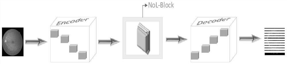
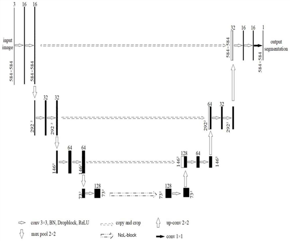
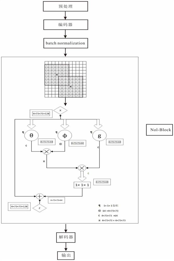
The invention provides a U-Net fundus retinal blood vessel image segmentation method based on improvement. The method comprises the following steps: acquiring a to-be-detected retinal blood vessel image; inputting the retinal blood vessel image into a trained improved U-Net network model for image segmentation; and outputting a retinal blood vessel image segmentation result. A common convolution block of the U-Net network is changed into a random discarded convolution block, so that image related features can be better extracted, and network over-fitting is effectively relieved; then, a NoL-block attention module is added to the bottom of a coding and decoding structure, and on the basis of not increasing the parameter quantity, the receptive field is expanded, and the correlation of pixel information is enhanced; the NoL-block is used as a component and is embedded into the U-Net network structure, so that the NoL-block can be used for directly capturing the spatial dependency relationship between pixels with distances in image segmentation in a forward propagation manner, and the blood vessel can be better positioned and segmented.
Description
technical field [0001] The invention belongs to the field of image segmentation, and in particular relates to an improved U-Net-based fundus retinal blood vessel image segmentation method and device. Background technique [0002] Segmentation of retinal vessels is important for the diagnosis and screening of retinal vascular diseases, including diabetes, ophthalmic diseases, and cardiovascular diseases. With the increasing number of visually impaired patients and the shortage of medical resources, especially the serious shortage of qualified ophthalmologists, ophthalmology patients often suffer from irreversible vision damage due to missing the best treatment period. The precise segmentation of fundus blood vessels can effectively assist ophthalmologists in their diagnosis and improve their work efficiency. [0003] With the continuous development of image processing technology, existing methods can be divided into unsupervised methods and supervised methods according to wh...
Claims
Login to View More Application Information
Patent Timeline
 IPC IPC(8): G06T7/00G06T7/11G06K9/62G06N3/04G06N3/08
CPCG06T7/0012G06T7/11G06N3/08G06T2207/10004G06T2207/20021G06T2207/20081G06T2207/20084G06T2207/30041G06T2207/30101G06N3/045G06F18/214Y02T10/40
Inventor韩静园王育坚宫浩栋
OwnerBEIJING UNION UNIVERSITY