
Cell detection method, device and equipment based on pathological image and storage medium

A technology of pathological images and cells, applied in the field of image processing, can solve the problem of occupying the time of the recognizer

Pending Publication Date: 2021-12-10
志诺维思(北京)基因科技有限公司
View PDF0 Cites 2 Cited by
  • Summary
  • Abstract
  • Description
  • Claims
  • Application Information

AI Technical Summary

Problems solved by technology

[0003] In the existing method, in order to obtain the number of different types of cells in the pathological image, it is necessary to accurately locate each cell in the pathological image and distinguish the cell type based on the identification method of human naked eyes. This method not only requires the recognizer to have rich correlation Experienced professionals, the entire identification process will take up a lot of time

Method used


Image

  • Cell detection method, device and equipment based on pathological image and storage medium
  • Cell detection method, device and equipment based on pathological image and storage medium
  • Cell detection method, device and equipment based on pathological image and storage medium

Examples


Embodiment 1

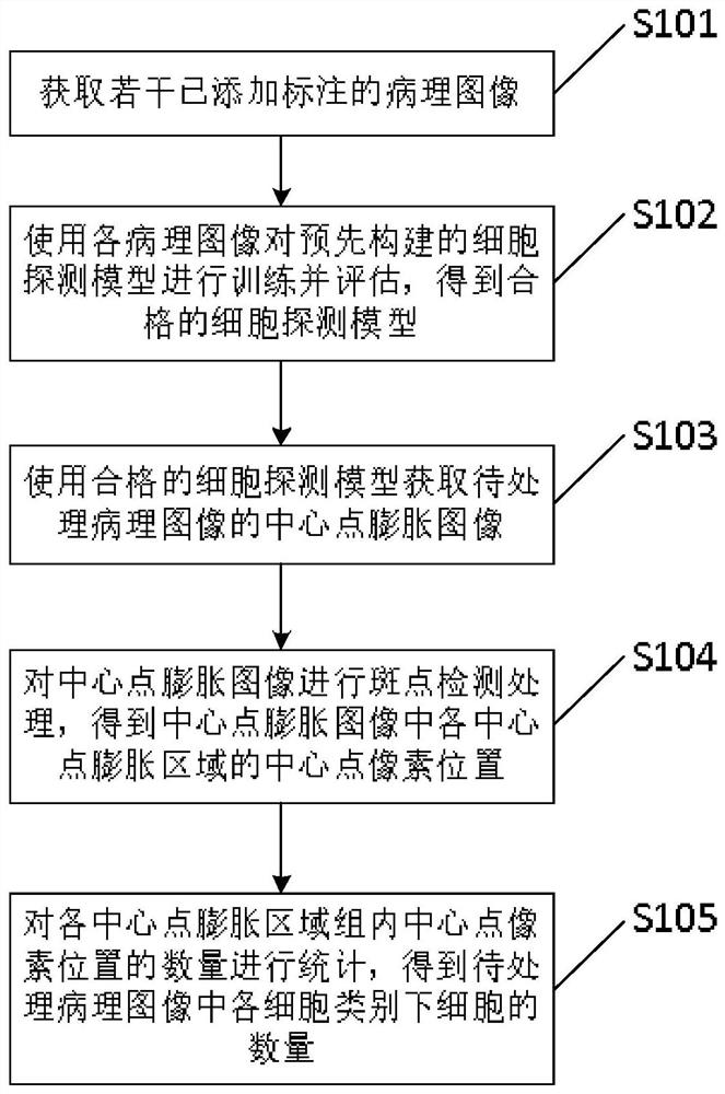
[0065] figure 1 It shows a flow chart of a pathological image-based cell detection method provided by Embodiment 1 of the present application, as shown in figure 1 As shown, the method includes the following steps:

[0066] Step S101: Obtain several pathological images with annotations added, wherein the annotations include an annotation of the cell center position and cell type of at least one cell in the pathological image.

[0067] Specifically, each pathological image includes at least one type of cell (that is, there is at least one cell category). In order to train and evaluate the pre-built cell detection model, it is necessary to obtain several pathological images with annotations (that is, pathological images with annotations). image), for each pathological image, the labeling on the pathological image includes the labeling of the cell center position of each cell in the pathological image and the labeling of the cell type of each cell, and the cell center position r...

Embodiment 2

[0123] Figure 9 A schematic structural diagram of a pathological image-based cell detection device provided in Embodiment 2 of the present application is shown, as shown in Figure 9 As shown, the cell detection device based on pathological images includes:

[0124] An acquisition module 501, configured to acquire a number of pathological images to which annotations have been added, wherein the annotations include an annotation of a cell center position and a cell type of at least one cell in the pathological image;

[0125] An execution module 502, configured to use each of the pathological images to train and evaluate a pre-built cell detection model to obtain a qualified cell detection model, wherein the pre-built cell detection model is a semantic segmentation model;

[0126] The detection module 503 is configured to use the qualified cell detection model to obtain a center point dilation image of the pathological image to be processed, wherein the center point expansion...

Embodiment 3

[0146] The embodiment of the present application also provides a computer device 600, Figure 10 It shows a schematic structural diagram of a computer device provided by Embodiment 3 of the present application, as shown in Figure 10 As shown, the device includes a memory 601, a processor 602, and a computer program stored in the memory 601 and operable on the processor 602, wherein, when the processor 602 executes the computer program, the above-mentioned pathological image-based cell detection method.

[0147] Specifically, the above-mentioned memory 601 and processor 602 can be general-purpose memory and processor, which are not specifically limited here. When the processor 602 runs the computer program stored in the memory 601, it can execute the above-mentioned cell detection method based on pathological images. It is beneficial to improve the positioning efficiency and counting efficiency of different types of cells while reducing the labor burden.


PUM


Abstract

The invention provides a cell detection method and device and equipment based on pathological images, and a storage medium, and the method comprises the steps of obtaining a plurality of pathological images with added labels; training and evaluating a pre-constructed cell detection model by using each pathological image to obtain a qualified cell detection model; acquiring a central point expansion image of the pathological image to be processed by using the qualified cell detection model; performing spot detection processing on the central point expansion image to obtain a central point pixel position of each central point expansion area in the central point expansion image; counting the number of central point pixel positions in each central point expansion region group to obtain the number of cells under each cell category in the pathological image to be processed. Through the method, the positioning efficiency and the counting efficiency of different types of cells are improved while the labor burden is reduced.

Description

technical field [0001] The present application relates to the technical field of image processing, in particular, to a pathological image-based cell detection method, device, equipment, and storage medium. Background technique [0002] Immunohistochemistry (IHC) is a method widely used in pathological diagnosis and guiding tumor treatment. It uses the chemical reaction method of antigen-antibody enzyme labeling to stain specific antigens in tissue cells, and the stained tissue The section is digitally scanned to obtain a pathological image, which reflects the cell morphology in the tissue and the expression of specific protein molecular markers, providing important information for doctors in clinical diagnosis, and guiding clinical tumor targeting and immunotherapy to formulate treatment strategies and solutions; and in order to facilitate doctors to accurately and quickly understand pathological images, it is necessary to first identify different types of cells and the numb...

Claims


Application Information

Patent Timeline
Patent Type & Authority Applications(China)
IPC IPC(8): G06T7/00G06T7/11G06T7/66G06T7/73G06T7/90G06K9/34
CPCG06T7/0012G06T7/11G06T7/66G06T7/73G06T7/90G06T2207/10024G06T2207/10056G06T2207/20021G06T2207/30024G06T2207/30242
Inventor 高楠楠王华刘昌灵张亚军凌少平
Owner 志诺维思(北京)基因科技有限公司
Learn More