Tissue defect closing instrument

A device and tissue technology, applied in the field of cardiac tissue defect closure device, can solve the problems of human tissue damage, poor occlusion stability, poor anchoring effect of occluder, etc., to reduce stimulation damage, reduce shear area, good The effect of biocompatibility

Active Publication Date: 2021-12-24
NINGBO DIOCHANGE MEDICAL TECH CO LTD
View PDF9 Cites 1 Cited by
  • Summary
  • Abstract
  • Description
  • Claims
  • Application Information

AI Technical Summary

Problems solved by technology

[0011] According to an embodiment in the application of the patent foramen ovale treatment operation, the present invention can provide a tissue defect closure device for patients with structural heart disease who need interventional treatment, which can solve the problem of using a sealing device during the interventional treatment operation. Some disadvantages caused by occluder treatment of patent foramen ovale surgery, such as poor occluder anchoring e

Method used


Image

  • Tissue defect closing instrument
  • Tissue defect closing instrument
  • Tissue defect closing instrument

Examples


Embodiment 1

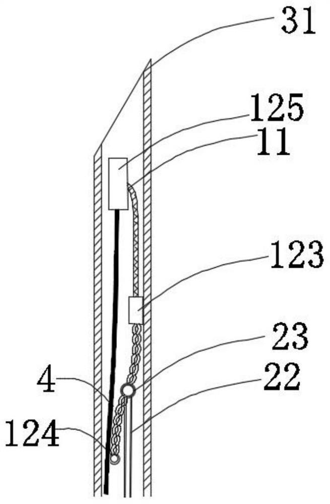
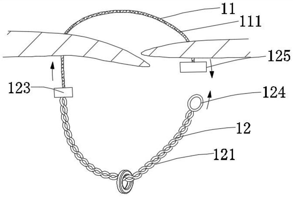
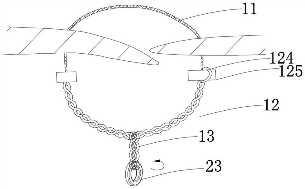
[0133] In this example, if Figure 1a~1c As shown, the closing device includes a closing unit and a release unit 13. The closing unit includes a connecting unit 11 and a locking unit 12 connected to the connecting unit 11. The locking unit 12 is located at the proximal end region of the connecting unit 11. The release unit 13 is located in the proximal region of the locking unit 12; the connecting unit 11 only includes one connecting piece 111; the locking unit 12 includes two locking pieces 121; the unlocking unit 13 is operated to drive the locking piece 121 to rotate , and the connecting unit 11 is tightened, the locking parts 121 are close to each other and realize self-locking locking, thereby realizing the closure of the tissue defect, and the closing unit part is connected with the tissue; the locking unit 12 and the release unit 13 are of an integrated structure, When the locking member 121 realizes self-locking and locking, the release unit 13 is further operated, so ...

Embodiment 2

[0160] The difference from Example 1 is:

[0161] In this example, if Figure 3b As shown, the closing unit includes a connecting unit 11 and one or more fixing members 16 located at the distal end region of the connecting unit 11; area, and connected with the connecting member 111, part or all of the fixing member 16 is attached or anchored to the inside of the target tissue or the outer surface of the distal end.

[0162] In this example, if Figure 3b As shown, when the connecting unit 11 located in the proximal region of the tissue defect is in an open state, the locking unit 12 and the connecting unit 11 are connected through the fixed connection structure 123 .

[0163] In this embodiment, the locking unit 12 includes two locking elements 121 , and each locking element 121 includes three wires.

[0164] In this embodiment, the fixing part 16 and the connecting part 111 are stretched in the tissue defect area, and the posture adjustment structure 14 adjusts the angle b...

Embodiment 3

[0190] The difference with embodiment one is:

[0191] In this example, if Figures 11a-11d As shown, the connecting unit 11 is a polymer wire, the locking unit 12 is a soft metal wire, and the locking unit 12 includes two locking parts 121, and each locking part 121 is connected by gluing, mechanical connection, welding, etc. On the connector and twisted with the connector 111 to form.

[0192] In this embodiment, the release unit 13 is operated so that the auxiliary breaking structure 8 drives the locking member 121 to rotate, and the locking members 121 are close to each other and realize self-locking locking, thereby realizing the closure of the tissue defect ; when the locking member 121 achieves self-locking and locking, the release unit 13 is further operated, and the auxiliary breaking structure 8 is further rotated, so that the auxiliary breaking structure 8 is broken and separated from the locking member 121, Such as Figures 11a-11d As shown, the connecting piece...


PUM


Abstract

The invention relates to the field of medical instruments, in particular to a tissue defect closing instrument. The closing instrument comprises a closing unit and a releasing unit, wherein the closing unit comprises a connecting unit and a locking unit connected with the connecting unit, the locking unit is located in the near-end area of the connecting unit, and the releasing unit is located in the near-end area of the locking unit; the connecting unit at least comprises one or more connecting pieces; the locking unit at least comprises one or more locking pieces; and the releasing unit is operated to drive the locking pieces to rotate, the connecting unit is tensioned, the locking pieces get close to each other and achieve self-locking, and then tissue defect closing is achieved. The locking unit and the releasing unit are of an integrated structure, and when the locking pieces achieve self-locking, the releasing unit is further operated, so that the releasing unit and the locking unit are broken and separated.

Description

technical field [0001] The invention belongs to the field of medical devices, in particular to a tissue defect closure device, in particular to a heart tissue defect closure device. Background technique [0002] Patent foramen ovale (Patent Foramen Oval, PFO) is the most common congenital heart abnormality in adults. In the normal population, about 1 person in 4 people has this disease. There are 1.5 million congenital heart disease patients in our country, and the annual prevalence rate of congenital heart disease is 6.78%. There are as many as 100,000-150,000 new-born children with congenital heart disease each year, of which simple patent foramen ovale accounts for about 10% of congenital heart disease. [0003] A patent foramen ovale is a common problem that can lead to potentially serious complications. In recent years, many studies have shown that there is a close relationship between patent foramen ovale and patients with stroke of unknown origin. For patients with...

Claims


Application Information

Patent Timeline
IPC IPC(8): A61B17/00
CPCA61B17/0057A61B2017/00663A61B2017/00575
Inventor 吕世文李彪邵烨
Owner NINGBO DIOCHANGE MEDICAL TECH CO LTD
Why Eureka
Try Eureka