
Method and device for detecting diseased tissue

A detection device and tissue technology, applied in the direction of measurement devices, test sample preparation, sampling, etc., can solve the problems of multiple equipment, complex detection process, and high work intensity of operators, so as to achieve a large amount of dissolution, avoid bending and wrinkling, The effect of simplifying the inspection process

Active Publication Date: 2022-05-20
HEILONGJIANG BAYI AGRICULTURAL UNIVERSITY
View PDF4 Cites 0 Cited by
  • Summary
  • Abstract
  • Description
  • Claims
  • Application Information

AI Technical Summary

Problems solved by technology

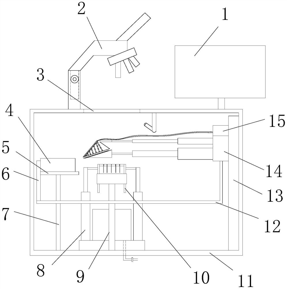
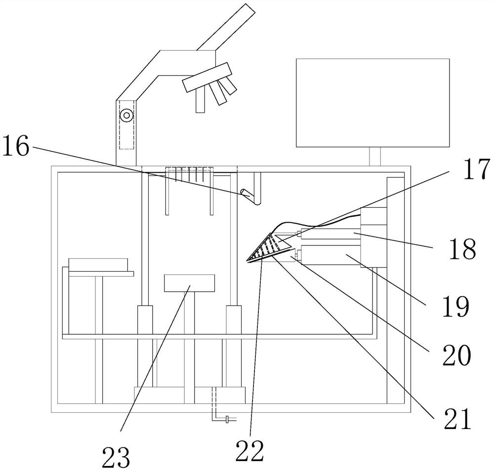
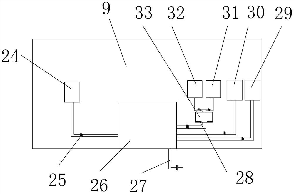
[0004] In view of the problems in the prior art that the detection process is complicated, the equipment involved is more, and the work intensity of the operator is high, the present invention provides a detection method and device for diseased tissue, the purpose of which is to reduce the use of equipment and save the occupied area of ​​equipment , to reduce the workload of the operator

Method used


Image

  • Method and device for detecting diseased tissue
  • Method and device for detecting diseased tissue
  • Method and device for detecting diseased tissue

Examples


Embodiment 1

[0056] A method for detecting diseased tissue, comprising the steps of:

[0057] Step S1, sampling and fixing: select a representative site for sampling, and the selected site should include the main lesion and the normal tissue connected to the lesion; fix the sample with a fixative immediately after sample collection;

[0058] Step S2, tissue trimming: using a cutting device to cut and trim the fixed sample, so that the sample initially assumes a regular shape;

[0059] Step S3, dehydration and transparency: use ethanol reagents to dehydrate the sample in gradient; after dehydration, use a transparent agent to perform transparent treatment on the sample;

[0060] Step S4, embedding in wax: put the transparent treated sample into melted paraffin and keep it at a constant temperature; pour the soaked sample and melted paraffin into the embedding box for cooling and solidification to form a wax block;

[0061] Step S5, slicing, dewaxing, staining and microscopic observation: B...


PUM

PropertyMeasurementUnit
thicknessaaaaaaaaaa

Abstract

The invention discloses a detection method and device for pathological tissue, which belongs to the technical field of tissue slice detection and solves the problems of complicated detection process, many equipments involved, and high work intensity of operators, which includes a detection method of tissue slices and The cutting device and microscopic imaging device used in the detection process, the cutting device is used for tissue trimming and wax block trimming, and the microscopic imaging device has the functions of sectioning, patching, dewaxing, staining and microscopic observation, Reduce the use of equipment, save the area occupied by equipment, and reduce the work intensity of operators.

Description

technical field [0001] The invention belongs to the technical field of tissue slice detection, in particular to a detection method and device for diseased tissue. Background technique [0002] Pathological examination has been widely used in clinical work and scientific research. In clinical practice, pathological examination of cadavers and surgical pathology are mainly carried out. The purpose of surgical pathological examination is, on the one hand, to clarify the diagnosis and verify the preoperative diagnosis, and improve the level of clinical diagnosis; on the other hand, after the diagnosis is confirmed, the next treatment plan can be determined and the prognosis can be estimated, thereby improving the level of clinical treatment. Through clinical and pathological analysis, a large amount of extremely valuable scientific research data can also be obtained. Pathological slices are a type of pathological specimens. Part of the diseased tissues or organs are treated wit...

Claims


Application Information

Patent Timeline
Patent Type & Authority Patents(China)
IPC IPC(8): G01N21/84G01N21/01G01N1/36G01N1/30G01N1/28
CPCG01N21/84G01N21/01G01N1/286G01N1/30G01N1/36G01N2001/2873G01N2021/0112G01N2001/302
Inventor 栾广宇原冬伟孙斌鲁文赓刘鹏
Owner HEILONGJIANG BAYI AGRICULTURAL UNIVERSITY
Learn More