Heart MRI segmentation method and system

A heart and segmentation model technology, applied in the field of image processing, can solve problems such as over-segmentation, and achieve the effect of improving segmentation accuracy and accuracy

Inactive Publication Date: 2022-01-07
UNIV OF ELECTRONIC SCI & TECH OF CHINA
View PDF0 Cites 0 Cited by
  • Summary
  • Abstract
  • Description
  • Claims
  • Application Information

AI Technical Summary

Problems solved by technology

[0007] Aiming at the above-mentioned deficiencies in the prior art, a cardiac MRI segmentation method and system provid

Method used


Image

  • Heart MRI segmentation method and system
  • Heart MRI segmentation method and system
  • Heart MRI segmentation method and system

Examples


Example Embodiment

[0060] Example 1

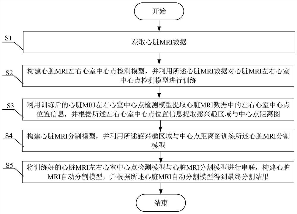
[0061] like figure 1 As shown, the present invention provides a cardiac MRI division method, which act is as follows:

[0062] S1, get heart MRI data;

[0063] In this embodiment, the short-axis heart MRI data disclosed on the network is collected and has an expert labeling result, such as the MICCAI 2013 heart MRI segmentation competition data set, which includes normal people and short axis heart movie MRI images, each The data contains a complete heartcy cycle of the subject.

[0064] In this embodiment, the data set is divided into three parts: training set, verification set and test set, and utilize the mean, maximum minimum normalization method and data amplification method (random shear, image Rotation, image contrast change, etc.) Treatment of the heart MRI data of the training set to obtain the database required for model training.

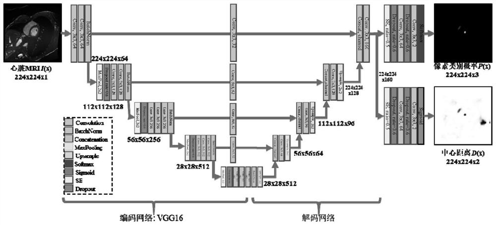
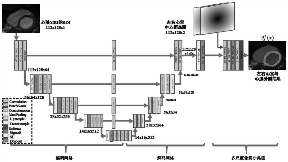
[0065] S2, construct a heart MRI left and right ventricular center point detection model, and use the heart MRI data to t...

Example Embodiment

[0099] Example 2

[0100] like Figure 4 As shown, the present invention provides a cardiac MRI division system, including:

[0101] Data acquisition module for obtaining cardiac MRI data;

[0102] Cardiac MRI left and right ventricular center point detection model build module, used to construct a heart MRI left-right ventricular center point detection model, and use the heart MRI data to train the center point detection model of the heart MRI left and right ventricular center points;

[0103] The extraction module is used to extract the left and right ventricular center points information in the heart MRI data using the trained heart MRI left and right ventricular center point detection model, and extract the region and the central point distance of the region according to the left and right ventricular center point position information;

[0104] Cardiac MRI Segmentation Model Construction Module for constructing a heart MRI segmentation model, and training the heart MRI segmenta...


PUM


Abstract

The invention provides a heart MRI segmentation method and system, and belongs to the technical field of image processing. According to the method, a two-stage full convolutional network model is adopted; the full convolutional network model in the first stage is used for detecting center points of left and right ventricles of the heart MRI, center point information is used for generating a heart MRI region-of-interest, so that the influence of surrounding similar tissues is reduced; meanwhile, center distance information of the left and right ventricles is generated by utilizing Euclidean distance transformation as prior information of the second-stage full convolutional network model; therefore, the segmentation precision of the left ventricle and the right ventricle and the cardiac muscle of the heart MRI is remarkably improved; meanwhile, the models of the first stage and the second stage are connected in series, the segmentation precision is effectively improved, and the over-segmentation and under-segmentation problems existing in heart MRI segmentation are effectively solved.

Description

Technical field [0001] The present invention belongs to the technical field of image processing, more particularly to a method and system for cardiac MRI segmentation. Background technique [0002] "China Cardiovascular Health and Disease Report 2019" that, as of 2019 the number of Chinese residents in cardiovascular disease prevalence as high as 330 million people, and the prevalence continues to rise in stages, to a heavy burden on the domestic economy. Since the heart cine MRI has a high resolution, non-invasive, radiation-free, multi-plane and other advantages, become the gold standard for diagnosis of cardiovascular disease. By manual segmentation of MRI anatomical structure of the heart (left and right ventricle), then extract ventricular function parameters (ventricular volumes, ejection fraction, stroke volume, cardiac wall thickness, and movement distance, etc.), cardiovascular disease may assist doctors precise the analysis to improve the diagnostic accuracy of the dise...

Claims


Application Information

Patent Timeline
IPC IPC(8): G06T7/00G06T7/11G06T7/62G06T7/73G06V10/764G06V10/82G06N3/04G06N3/08
CPCG06T7/0012G06T7/11G06T7/62G06T7/73G06N3/08G06T2207/10088G06T2207/20016G06T2207/30048G06N3/047G06N3/048G06N3/045G06F18/2415
Inventor 李纯明谢李鹏
Owner UNIV OF ELECTRONIC SCI & TECH OF CHINA
Why Eureka
Try Eureka