Full-automatic quantitative analysis method for perinuclear lysosome distribution

A quantitative analysis, lysosome technology, applied in the field of cell analysis, can solve problems such as difficult to achieve high-throughput big data analysis, and achieve the effect of computational robustness

Pending Publication Date: 2022-01-14
北京中科与点科技中心(有限合伙)
View PDF0 Cites 0 Cited by
  • Summary
  • Abstract
  • Description
  • Claims
  • Application Information

AI Technical Summary

Problems solved by technology

Most of the existing work manually specifies and divides these areas, which is difficult to achieve high-throughput big data analysis

Method used


Image

  • Full-automatic quantitative analysis method for perinuclear lysosome distribution
  • Full-automatic quantitative analysis method for perinuclear lysosome distribution
  • Full-automatic quantitative analysis method for perinuclear lysosome distribution

Examples


no. 1 example

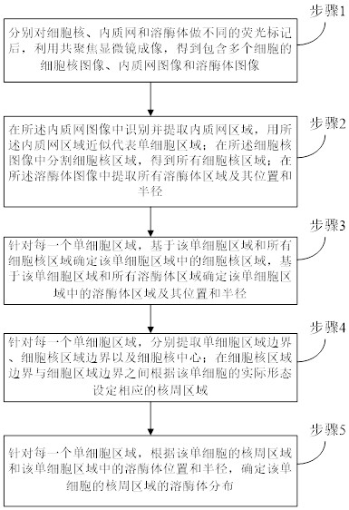
[0040] In the first embodiment of the present invention, a nuclear cycosocidizer distribution automatic quantitative analysis method, such as figure 1 As shown, the following specific steps are included:

[0041] Step 1, after different fluorescence markers of the nucleus, the nucleus microscopy is used to obtain a nucleus image, an endometric web image, and a lysosomas image including a plurality of cells.

[0042] Step 2, identify and extract the endoplasmic network area in the inner textone web image, and use the inner texture network area to represent a single cell region; in the nuclear image, the nucleus is divided into all nuclear region; All lysosomas are extracted in the lysosomal image and its position and the radius thereof;

[0043]Alternatively, in the step 2, a depth learning-based instance division model MASKRCNN is identified and extracted in the endoplasmic web region, and the endogenic network region is used to approximate a single cell region;

[0044] In the st...

no. 2 example

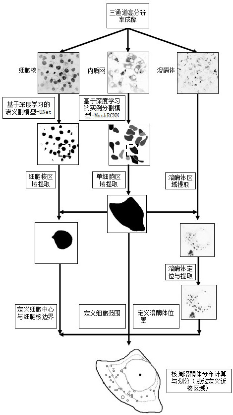
[0066] In the second embodiment of the present invention, the present embodiment is incorporated herein by reference. Figure 2 ~ 3 An application example of the present invention is described.

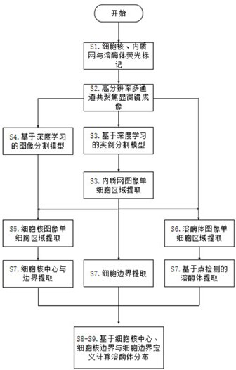
[0067] like image 3 As shown, the nuclear cycosome distribution automatic quantitative analysis method according to the embodiment of the present invention, including the following specific steps:

[0068] Step S1: Different fluorescent markers are made to the nucleus, endometric webs and lysosomes.

[0069] Step S2: Through high-resolution multi-channel copolymerization microscopy, it is obtained from nuclear, endometric webs and lysosomas, each image containing multiple cells, generally more than 10, such as figure 2 Indicated.

[0070] Step S3: By using the inner texture network, the characteristics of the entire cell region are occupied by the internal quality web image. Use the depth learning-based instance division model Maskrcnn to identify and extract the endometric network area, a...

no. 3 example

[0081] According to a third embodiment of the present invention, the flow of nuclear cyclic exclusive distribution automatic quantitative analysis method is the same, and the second embodiment is the same, and the present embodiment can be used in the project implementation. The general purpose hardware platform is implemented, of course, can also pass hardware, but in many cases, the former is better implementation. Based on this, the method of the present invention can be embodied in the form of a computer software product, which is stored in a storage medium (such as ROM / RAM, disk, disc), including several instructions to make one The method described in the embodiment of the present invention is performed.


PUM


Abstract

The invention provides a full-automatic quantitative analysis method for perinuclear lysosome distribution, which comprises the following steps: identifying and extracting an endoplasmic reticulum region in an endoplasmic reticulum image, and approximately representing a single cell region by using the endoplasmic reticulum region; segmenting cell nucleus regions in a cell nucleus image to obtain all cell nucleus regions; extracting all lysosome regions from a lysosome image; aiming at each single cell region, determining a cell nucleus region and a lysosome region in the single cell region; setting a corresponding perikaryotic region according to the actual form of the single cell; and determining lysosome distribution of the perinuclear region of the single cell according to the perinuclear region of the single cell and the position and radius of the lysosome in the single cell region. According to the method, an automatic analysis process is designed based on a deep learning technology, accurate perinuclear lysosome distribution quantitative analysis is realized, the problem of manual calculation at present is solved, and high-throughput big data analysis becomes possible.

Description

Technical field [0001] The present invention relates to the field of cell analysis, and more particularly to a nuclear cycle-free distribution automatic quantitative analysis method. Background technique [0002] The lysosomal distribution in the cells exhibits a certain state, such as the lysosomes of aging cells generally aggregate to the nuclear circumference region. In the relevant field, there is still a lack of effective means to achieve accurate nuclear cycosome distribution quantitative analysis, and its technical difficulties are precisely defining the cell center and cell boundary, and accurately engraving nuclear asterities, such as quantization of the vision of the farming area. Most of the existing work is manually designated and divided into these areas, it is difficult to achieve high-throughput large data analysis. How to automate the distribution of nuclear cyclic exclusive distributions by means of a computer is a technical issue that is urgent to solve the need...

Claims


Application Information

Patent Timeline
IPC IPC(8): G06T7/00G06T7/11G06T7/73G06T7/62G06N3/04G06N3/08
CPCG06T7/0012G06T7/11G06T7/73G06T7/62G06N3/08G06T2207/10056G06T2207/30024G06T2207/20081G06N3/045
Inventor 周东张朝栋王育雄刘琳琳
Owner 北京中科与点科技中心(有限合伙)
Why Eureka
Try Eureka