
Thyroid gland detection device

A detection device, thyroid technology, applied in the direction of organ motion/change detection, image data processing, diagnosis, etc., can solve problems such as limiting the wide application of ultrasound, and achieve the effect of facilitating subsequent analysis

Pending Publication Date: 2022-01-18
什维新智医疗科技(上海)有限公司
View PDF0 Cites 0 Cited by
  • Summary
  • Abstract
  • Description
  • Claims
  • Application Information

AI Technical Summary

Problems solved by technology

However, in current clinical practice, the reading of ultrasound image features largely depends on the subjective or semi-subjective analysis of images by sonographers, which limits the wide application of ultrasound.
However, there are no studies on quantitative feature analysis of TI-RADS to improve the diagnostic performance of thyroid cancer classification.

Method used


Image

  • Thyroid gland detection device
  • Thyroid gland detection device
  • Thyroid gland detection device

Examples


Embodiment Construction

[0024] Below in conjunction with specific embodiment, further illustrate the present invention. It should be understood that these examples are only used to illustrate the present invention and are not intended to limit the scope of the present invention. In addition, it should be understood that after reading the teachings of the present invention, those skilled in the art can make various changes or modifications to the present invention, and these equivalent forms also fall within the scope defined by the appended claims of the present application.

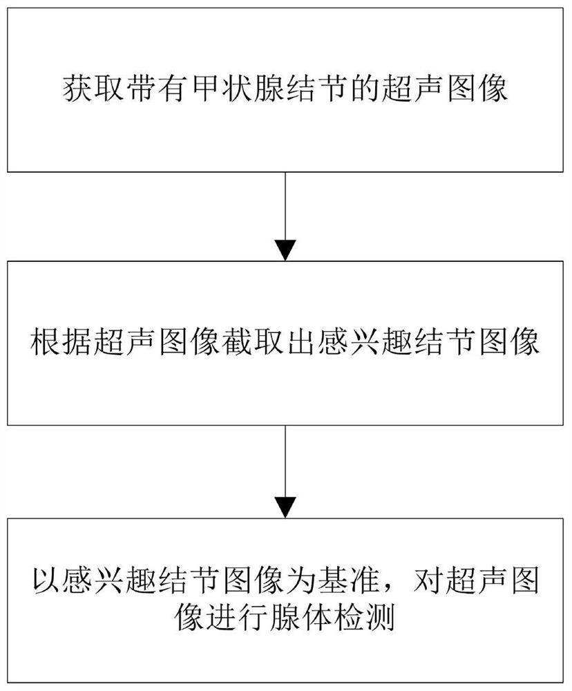
[0025] Embodiments of the present invention relate to a thyroid gland detection device, please refer to figure 1 ,include:

[0026] Image acquisition module: used to acquire ultrasound images with thyroid nodules;

[0027] Ultrasonic image preprocessing module: used to filter the ultrasonic image through an adaptive median filter to remove speckle noise in the ultrasonic image; filtering for enhancing the contrast and edges ...


PUM


Abstract

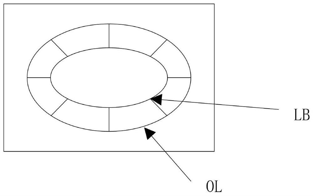
The invention relates to a thyroid gland detection device. The thyroid gland detection device comprises an image acquisition module which is used for acquiring an ultrasonic image with thyroid nodules; a nodule-of-interest image extraction module which is used for intercepting the thyroid nodule boundary of the ultrasonic image by selecting a plurality of coordinate points of interest to obtain a nodule-of-interest image; and a thyroid gland detection module which is used for carrying out gland detection on the ultrasonic image by taking the nodule in the nodule-of-interest image as a reference. The thyroid gland can be effectively detected.

Description

technical field [0001] The invention relates to the technical field of auxiliary medical diagnosis, in particular to a thyroid gland detection device. Background technique [0002] Thyroid cancer is one of the most common cancers in women worldwide, with women three times more likely than men. In 2018, one in 20 cancers diagnosed in women was thyroid cancer. Ultrasound imaging is a non-invasive, radiation-free, and low-cost tumor diagnostic technique. However, identifying thyroid nodules and detecting tumor signs from ultrasound images is a challenging task due to the low quality of ultrasound images. [0003] In general, the growth and development of a malignant tumor can be reflected by its shape, appearance, texture, composition, and many other factors. As an effective tool, gray-scale ultrasound (US) images can visualize these factors, helping doctors to better observe and understand the performance of pathological sites. However, in current clinical practice, the re...

Claims


Application Information

Patent Timeline
Patent Type & Authority Applications(China)
IPC IPC(8): A61B8/08G06T5/00G06T5/20G06T7/00G06T7/73
CPCA61B8/085A61B8/5215A61B8/5269G06T7/0012G06T7/73G06T5/20G06T2207/10132G06T2207/20032G06T2207/20104G06T2207/20192G06T5/90G06T5/70
Inventor 于明
Owner 什维新智医疗科技(上海)有限公司
Learn More