DR normal position chest radiograph clavicle segmentation method and device in quality control, processing equipment and storage medium

A clavicle and chest X-ray technology, applied in the field of DR image recognition, can solve the problems of slow clavicle positioning and low efficiency of image quality control and evaluation, achieve fast segmentation, improve the efficiency of image quality control and evaluation, and save the use of finding clavicles the effect of time

Pending Publication Date: 2022-01-28
心医国际数字医疗系统(大连)有限公司
View PDF0 Cites 1 Cited by
  • Summary
  • Abstract
  • Description
  • Claims
  • Application Information

AI Technical Summary

Problems solved by technology

[0003] In view of this, the object of the present invention is to provide a method, device, processing equipment and storage medium for segmenting the clavicle in DR anteroposterior chest radiographs in quality control, so as to solve the problem of slow clavicle positioning speed and image quality control evaluation work efficiency in the prior art. low problem

Method used


Image

  • DR normal position chest radiograph clavicle segmentation method and device in quality control, processing equipment and storage medium
  • DR normal position chest radiograph clavicle segmentation method and device in quality control, processing equipment and storage medium
  • DR normal position chest radiograph clavicle segmentation method and device in quality control, processing equipment and storage medium

Examples


Embodiment 1

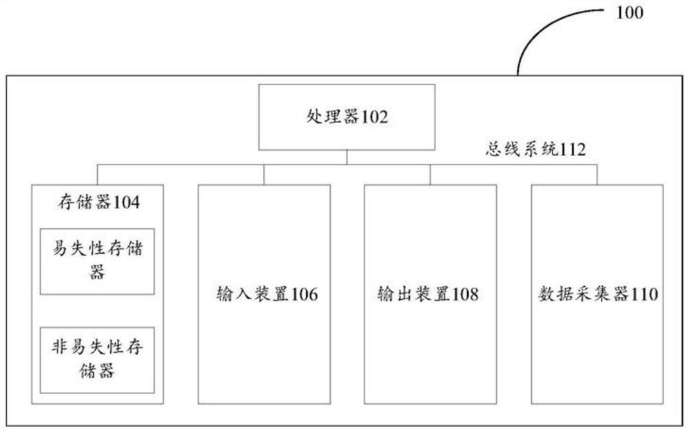
[0047] First, refer to figure 1 The processing device 100 for implementing the embodiments of the present invention will be described, and the processing device can be used to run the methods of the various embodiments of the present invention.

[0048] Such as figure 1 As shown, the processing device 100 includes one or more processors 102, one or more memories 104, an input device 106, an output device 108, and a data collector 110, and these components are connected through a bus system 112 and / or other forms ( not shown) interconnection. It should be noted that figure 1The components and structure of the processing device 100 shown are only exemplary, not limiting, and the processing device may also have other components and structures as required.

[0049] The processor 102 may be implemented in at least one hardware form of a digital signal processor (DSP), a field programmable gate array (FPGA), a programmable logic array (PLA) and an ASIC (Application Specific Integ...

Embodiment 2

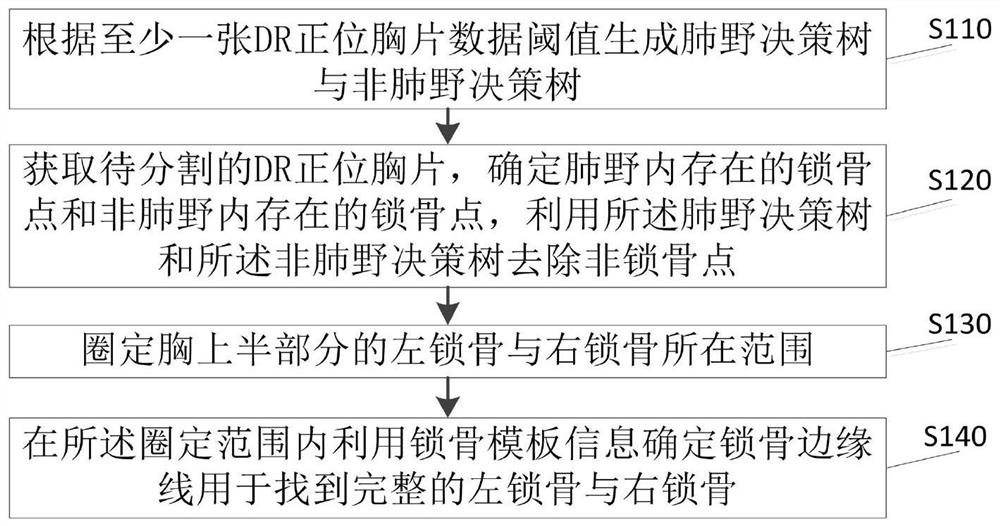
[0056] see figure 2 The shown method for segmenting the clavicle of DR anteroposterior chest radiograph in quality control is applied to a mobile terminal, and the method can be executed by the processing device provided in the foregoing embodiment. The method specifically includes the following steps:

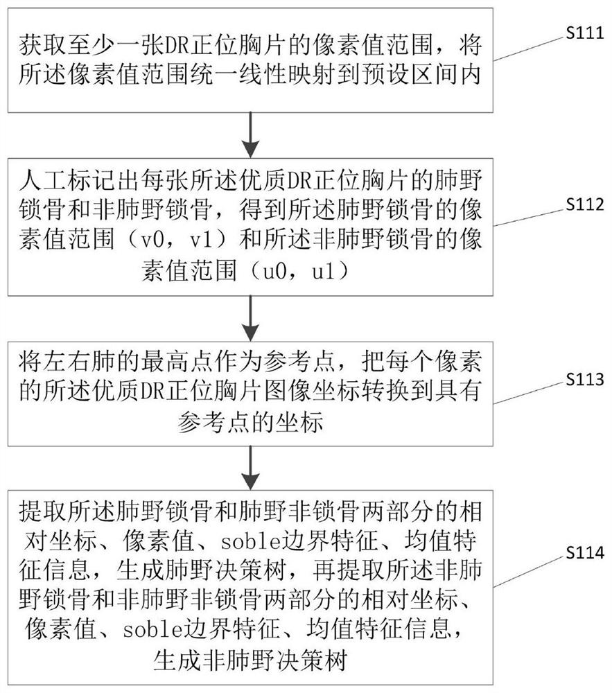
[0057] S110. Generate a lung field decision tree and a non-lung field decision tree according to the threshold value of at least one DR frontal chest radiograph data;

[0058] Among them, at least one DR chest radiograph is selected from the database and has been manually evaluated as high-quality DR chest radiographs, and the number of them is not less than 100. Calculate a minimum bounding box R(x1, y1, x2, y2) including the clavicle.

[0059] S120. Obtain the DR frontal chest radiograph to be segmented, determine the clavicular points existing in the lung field and the clavicular points existing in the non-lung field, and use the lung field decision tree and the non-lung ...

Embodiment 3

[0090] Regarding the method for segmenting clavicles of DR anteroposterior chest radiographs in quality control provided in Embodiment 2, an embodiment of the present invention provides a device for segmenting clavicles of DR anteroposterior chest radiographs in quality control, which is applied to mobile terminals, see Image 6 A structural block diagram of a clavicle segmentation device for DR anteroposterior chest X-ray in quality control shown, including:

[0091]A generating module 610, configured to generate a lung field decision tree and a non-lung field decision tree according to at least one DR frontal chest radiograph data threshold;

[0092] The distinguishing module 620 is used to obtain the DR frontal chest radiograph to be segmented, determine the clavicle points existing in the lung field and the clavicle points in the non-lung field, and use the lung field decision tree and the non-lung field decision tree to remove the non-clavicle point;

[0093] The delinea...


PUM


Abstract

The invention discloses a DR normal position chest radiograph clavicle segmentation method and device in quality control, processing equipment and a storage medium, and relates to the technical field of DR image recognition. The segmentation method comprises the following steps: generating a lung field decision tree and a non-lung field decision tree according to a data threshold value of at least one DR normal position chest radiograph; obtaining a to-be-segmented DR normal position chest radiograph, determining clavicle points existing in a lung field and clavicle points existing in a non-lung field, and removing non-clavicle points by using the lung field decision tree and the non-lung field decision tree; delineating the range of the left clavicle and the right clavicle of the upper half part of the chest; and determining a clavicle edge line in the delineated range by utilizing clavicle template information so as to find a complete left clavicle and a complete right clavicle. Manual operation is not needed, the clavicle is completely and automatically segmented, the segmentation accuracy can be guaranteed, the segmentation speed is high, and the vertebra is subsequently positioned by using the clavicle position and named. In the whole process, the time for finding the clavicle is greatly saved, and the working efficiency of image quality control evaluation is obviously improved.

Description

technical field [0001] The present invention relates to the technical field of DR image recognition, in particular to a method, device, processing equipment and storage medium for segmenting clavicles of DR anteroposterior chest radiographs in quality control. Background technique [0002] At present, with the continuous development of my country's medical industry, various inspection and treatment equipment are also constantly improving. In various clinical disciplines, imaging technology occupies an important position in diagnostic examinations, and the quality of imaging directly affects diagnostic examinations. The accuracy of the results will affect the accuracy of the treatment measures. Therefore, doing a good job in image quality control is one of the key clinical tasks of each hospital. In addition, the rapid development of regional images and cloud images this year has increased the requirements for image quality. Due to the huge number of images, manual quality con...

Claims


Application Information

Patent Timeline
Patent Type & Authority Applications(China)
IPC IPC(8): G06T7/12G06T7/13G06T7/136
CPCG06T7/12G06T7/136G06T7/13G06T2207/10124G06T2207/30008
Inventor 刘龙
Owner 心医国际数字医疗系统(大连)有限公司
Why Eureka
Try Eureka