Image exposure imaging method, imaging device and readable storage medium

An imaging method and image technology, applied in the field of medical devices, can solve the problems such as the inability to improve the image resolution of the tissue or organ of interest to the doctor, affecting the doctor's observation and judgment of the patient's tissue, and reducing the operating efficiency of the chief surgeon. Judging, improving the accuracy of surgery, and the effect of small hardware resource allocation

Pending Publication Date: 2022-02-08
SHANGHAI MICROPORT MEDBOT (GRP) CO LTD
View PDF0 Cites 2 Cited by
  • Summary
  • Abstract
  • Description
  • Claims
  • Application Information

AI Technical Summary

Problems solved by technology

At present, the commonly used exposure methods of endoscope systems on the market are ordinary automatic exposure and drone interaction. When the doctor is interested in an image with overexposure or underexposure, the status quo of the image cannot be changed.
Specifically, the endoscope automatic exposure algorithm is commonly used to expose the image globally. When the backlight and light-facing areas appear in the image at the same time, it will affect the doctor's further observation and judgment of the patient's tissue, and affect the doctor's further observation of the tissue or organ of interest. Reduced surgical precision
[0004] Moreover, most of the existing endoscopic systems use the mouse to realize the interaction between the doctor and the image, such as digital zoom; this method not only has cumbersome operation steps, but also reduces the doctor’s operation efficiency and increases the doctor’s workload, and cannot improve the doctor’s interest in tissues or images. Organ image resolution
In addition, the commonly used endoscopic digital zoom method often requires the surgeon to click on the area of ​​interest with the mouse, which is complicated to operate and reduces the efficiency of the surgeon.

Method used


Image

  • Image exposure imaging method, imaging device and readable storage medium
  • Image exposure imaging method, imaging device and readable storage medium
  • Image exposure imaging method, imaging device and readable storage medium

Examples


Embodiment Construction

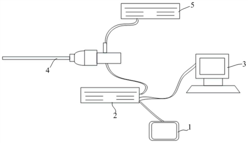
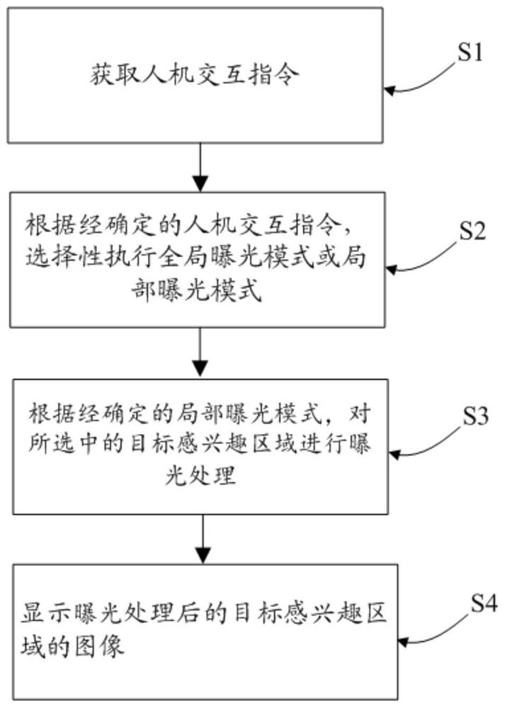
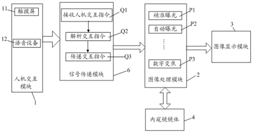
[0078] In order to make the purpose, advantages and features of the present invention clearer, the present invention will be further described in detail below in conjunction with the accompanying drawings and specific embodiments. It should be noted that the drawings are all in very simplified form and not drawn to scale, and are only used to facilitate and clearly assist the purpose of illustrating the embodiments of the present invention. In addition, the structures shown in the drawings are often a part of the actual structures. In particular, each drawing needs to display different emphases, and sometimes uses different scales.

[0079] As used in the present invention, the singular forms "a", "an" and "the" include plural objects, the term "or" is usually used in the sense of including "and / or", and the term "several" Usually, the term "at least one" is used in the meaning of "at least one", and the term "at least two" is usually used in the meaning of "two or more". In ...


PUM


Abstract

The invention relates to an image exposure imaging method, an imaging device and a readable storage medium. The image exposure imaging method comprises the following steps: providing a local exposure mode; and according to a determined man-machine interaction instruction, executing the local exposure mode to perform exposure processing on a selected target region of interest, and then displaying an image of the target region of interest after the exposure processing. Through the configuration, a user can obtain a good man-machine interaction mode, and the operation efficiency is improved; and a region of interest can be accurately exposed according to a man-machine interaction instruction, so that a clearer and more accurate view is provided for a doctor, the operation accuracy is improved, and harm of an operation to a patient is relieved.

Description

technical field [0001] The invention relates to the field of medical devices, in particular to an image exposure and imaging method, an imaging device and a readable storage medium. Background technique [0002] The endoscope enters the human body through the natural orifice of the human body or through a small incision made through surgery. When in use, the endoscope is introduced into the pre-examined organ, and the changes in the relevant parts can be directly observed. The quality of the image directly affects the use effect of the endoscope, and also marks the development level of the endoscope technology. [0003] Due to the narrow space inside the human body and limited light source conditions, the imaging quality of the endoscope system is seriously affected. Therefore, the quality of the exposure method plays a decisive role in the image quality. At present, the commonly used exposure methods of endoscope systems on the market are ordinary automatic exposure and ...

Claims


Application Information

Patent Timeline
Patent Type & Authority Applications(China)
IPC IPC(8): H04N5/232H04N5/243A61B1/00A61B1/045
CPCA61B1/00039A61B1/045A61B1/00163H04N23/67H04N23/62H04N23/611H04N23/76
Inventor 张莉何超
Owner SHANGHAI MICROPORT MEDBOT (GRP) CO LTD
Why Eureka
Try Eureka