Naked eye 3D spine microendoscope system

A 3D, naked-eye technology, applied in the fields of endoscopy, laparoscopy, medical science, etc., can solve the problems of difficult hand-eye coordination, loss of depth sense, poor visualization experience, etc., to prevent human eye tracking deviation and recognition errors. , Reduce the probability of crosstalk, and save the effect of target locking speed

Pending Publication Date: 2022-02-18
付强
View PDF0 Cites 0 Cited by
  • Summary
  • Abstract
  • Description
  • Claims
  • Application Information

AI Technical Summary

Problems solved by technology

Conventional spinal endoscopy is a two-dimensional display endoscope. Doctors lack stereo vision, loss of depth perception, and difficulty in hand-eye coordination, which increases the difficulty of surgery and increases the risk of nerve injury. Therefore, spinal endoscopic surgery usually requires long learning curve
[0003] Currently, common 2D spinal surgery endoscopic systems and polarized 3D surgical endoscopic systems are commonly used (auxiliary lenses are required to achieve 3D viewing effects). The spine endoscopes used in clinical and market applications are all 2D imaging. Its imaging quality such as clarity and resolution needs to be improved, which may easily cause loss of details, and lack of stereoscopic display effect, which may make it difficult to accurately locate during operation, insufficient restoration of picture fidelity, and poor visual experience may cause operational errors , increased risk of nerve damage
The existing 2D system imaging is not three-dimensional, and cannot accurately determine the spatial position of the anatomical structure, which increases operational risks; the existing 3D system needs to wear a special viewing device, which is not convenient enough, and the imaging is not clear enough, which is easy to cause dizziness and fatigue
Other glasses-free 3D display devices on the market are mostly dual-camera eye tracking or marker-type eye tracking, which requires very high processing technology for the camera, heavy equipment, cumbersome calibration methods, and high mass production costs.

Method used


Image

  • Naked eye 3D spine microendoscope system
  • Naked eye 3D spine microendoscope system
  • Naked eye 3D spine microendoscope system

Examples


Embodiment Construction

[0037] The present invention will be described in detail below in conjunction with the accompanying drawings and specific embodiments. The features such as component models, material names, connection structures, control methods, algorithms, etc. that are not clearly stated in this technical solution are regarded as common technical features disclosed in the prior art.

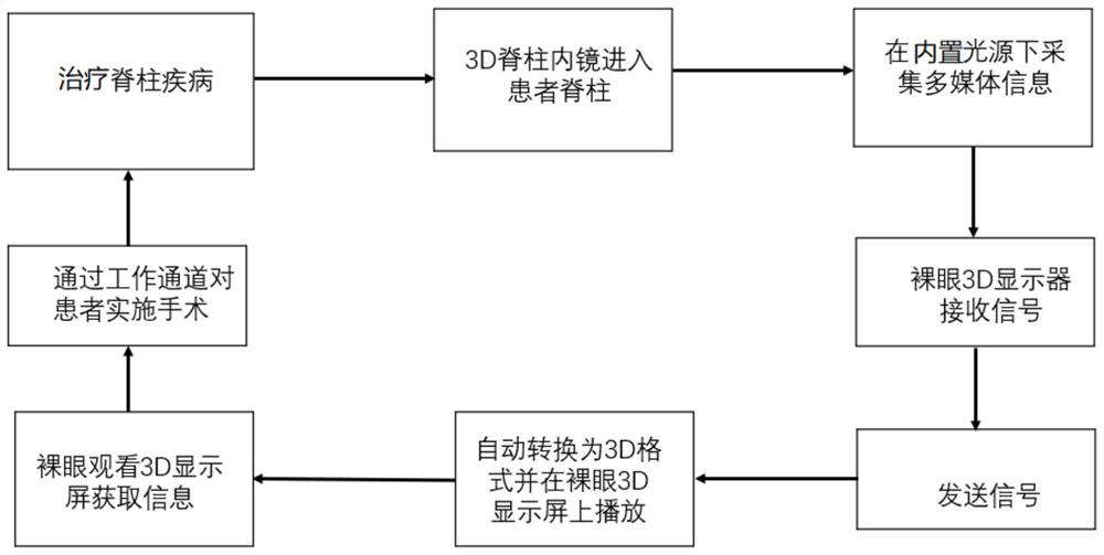
[0038] Starting from clinical pain points and actual needs, this device adopts a special 3D camera and naked-eye 3D display subsystem. During the treatment of patients with spinal diseases, the user can clearly see the 3D images of the patient's spine in real time, and can observe More details can prevent fatigue and dizziness caused by wearing auxiliary equipment for a long time. When performing surgery on the spine, it can improve the safety of the operation and achieve better surgical results. In addition, in terms of product design, a new type of single-camera eye tracking is adopted, which relies on human...


PUM


Abstract

The invention relates to a naked eye 3D spine microendoscope system, which comprises a 3D endoscope, a 3D image processor assembly and a naked eye 3D display device. The 3D endoscope comprises a 3D endoscope body and an endoscope lens connected with the 3D endoscope body. The 3D image processor assembly comprises a first FPGA (Field Programmable Gate Array) and a second FPGA which are electrically connected with each other. The first FPGA is electrically connected with the endoscope lens and converts each frame of image into a frame of three-dimensional image in a left-right eye format, and the second FPGA converts the three-dimensional image in the left-right eye format into a mixed three-dimensional image of optical parameters of the naked eye 3D display. The naked-eye 3D display device naturally splits the mixed stereoscopic image of the optical parameters of the naked-eye 3D display through a grating. Compared with the prior art, a special 3D camera and a naked eye 3D display subsystem are adopted, a user can clearly see 3D images in the spine of a patient in real time and get more details, and fatigue and dizziness caused by long-term wearing of auxiliary facilities are prevented.

Description

technical field [0001] The invention relates to the field of medical instruments, in particular to a naked-eye 3D spinal microendoscope system. Background technique [0002] Spinal endoscopic surgery is usually done in a small wound, the instruments are usually very long, and the doctor lacks tactile feedback, which makes the visual feedback of the spinal endoscopist particularly important during the operation, and compared with other endoscopic operation scenarios, the spinal structure is complex , There are many bone and nerve components, and a variety of organizational structures. It was previously considered a contraindication area for endoscopy. The space for spinal endoscopy is extremely small, and there are requirements for size. Conventional spinal endoscopy is a two-dimensional display endoscope. Doctors lack stereo vision, loss of depth perception, and difficulty in hand-eye coordination, which increases the difficulty of surgery and increases the risk of nerve inj...

Claims


Application Information

Patent Timeline
Patent Type & Authority Applications(China)
IPC IPC(8): A61B1/317A61B1/04
CPCA61B1/317A61B1/00195A61B1/04
Inventor 付强郭松晏美俊宋晓燕
Owner 付强
Why Eureka
Try Eureka