
Lens mount, distal module, endoscope and method

A technology of lens holder and distal end, which is applied in the field of endoscopy, can solve the problems of high difficulty, damage of lighting unit and camera unit, etc., and achieve the effects of improving quality, convenient loading, convenient loading and positioning

Active Publication Date: 2022-03-01
HUNAN VATHIN MEDICAL INSTR CO LTD
View PDF4 Cites 0 Cited by
  • Summary
  • Abstract
  • Description
  • Claims
  • Application Information

AI Technical Summary

Problems solved by technology

And when installing the lighting unit and camera unit, they are inserted from the rear end to the front end. In such a small space, it is very difficult to assemble through the rear end insertion, and it is easy to cause the lighting unit and camera unit to be damaged during assembly. damage

Method used


Image

  • Lens mount, distal module, endoscope and method
  • Lens mount, distal module, endoscope and method
  • Lens mount, distal module, endoscope and method

Examples


Embodiment 1

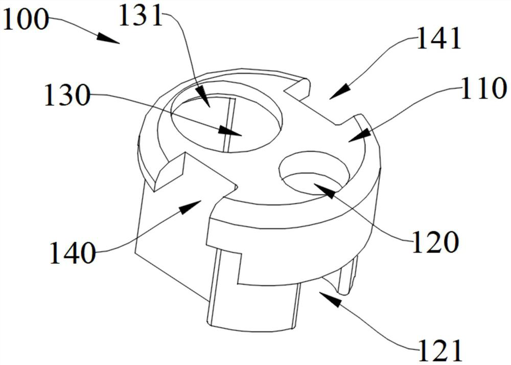
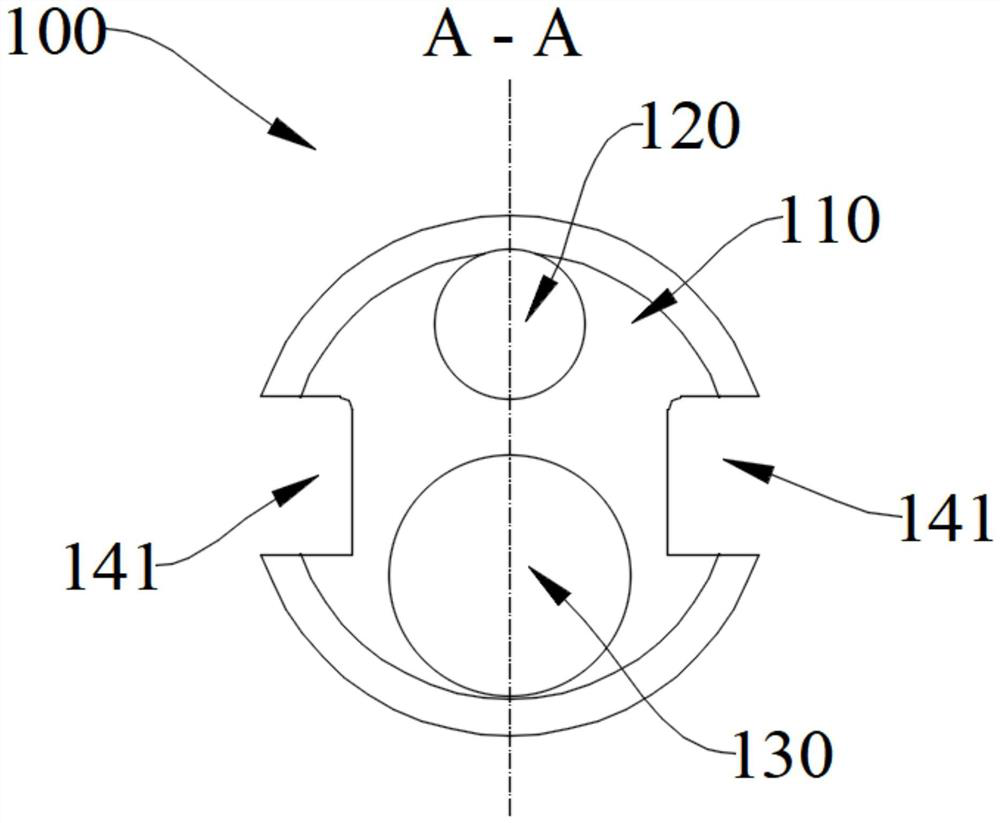
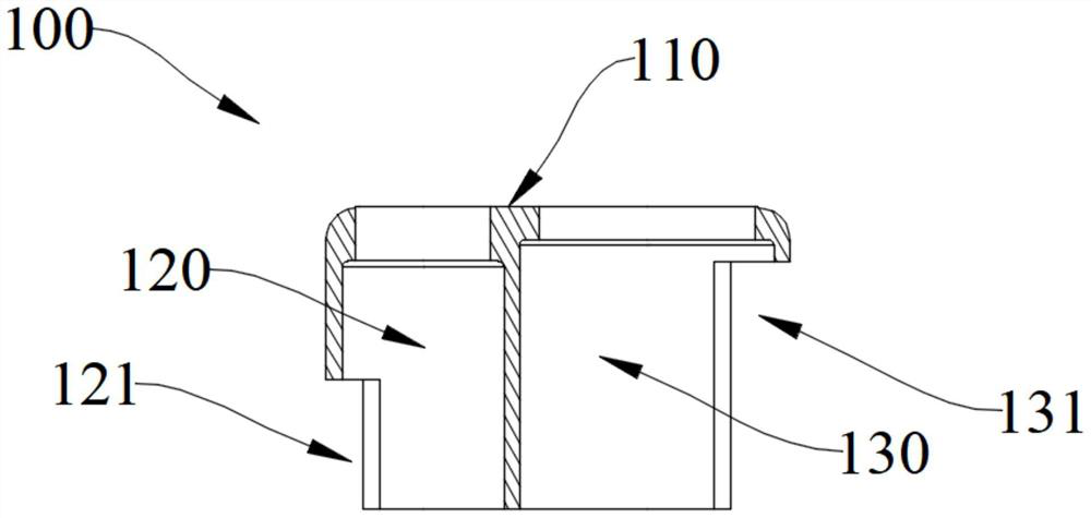
[0044]Embodiment 1 of the present invention provides a lens mount 100, a camera module housing portion is provided along the axial direction of the lens mount body 110, and the camera module housing portion includes a camera working channel 130 and an illumination working channel 140;

[0045] The side wall of the lens holder body 110 is provided with an opening of the camera module, and the opening of the camera module includes a first opening 131 and a second opening 141 adjacent to each other, and the first opening 131 and the second opening 141 are adjacent to each other. The camera working channel 130 communicates, the second opening 141 communicates with the lighting working channel 140 , the first opening 131 is used for loading the camera module, and the second opening 141 is used for installing the lighting module.

[0046] In this embodiment, there is no special limitation on the material of the lens holder body 110 , which may be a transparent material or an opaque m...

Embodiment 2

[0059] Embodiment 2 of the present invention provides a remote module 200, including a camera module 210 and the lens holder 100, the camera module 210 is at least partly accommodated in the camera module accommodating portion;

[0060] The camera module 210 includes a camera unit 211, a light emitting unit 212 and a supporting unit 213, the camera unit 211 and the light emitting unit 212 are arranged on the same side of the supporting unit 213, the camera unit 211, the light emitting unit The unit 212 is integrated with the support unit 213 .

[0061] The camera module is integrated, and the integrated camera module is installed into the lens holder body from the side, which realizes efficient assembly while avoiding damage, and effectively improves the success rate and assembly efficiency of the remote module 200 assembly.

[0062] Further, a sleeve 220 is also included, and the sleeve 220 is sleeved on the outer peripheral wall of the lens mount 100 . The sleeve 220 is use...

Embodiment 3

[0067] Embodiment 3 of the present invention provides an endoscope, including a handle portion and an insertion portion, the insertion portion includes a curved portion 310 and the distal module 200 , and the curved portion 310 is fixedly connected to the distal module 200 .

[0068] The handle part and the insertion part composed of the bending part 310 and the distal module 200 form an endoscope for detection of internal organs. At the same time, an operation tube 320 may also be provided in the endoscope, and the operation tube 320 may be used for sampling or medium circulation, so as to achieve the purpose of sampling or treatment.


PUM


Abstract

The invention is applicable to the technical field of endoscopes, and provides a lens mount, a far-end module, an endoscope and a method, the lens mount is provided with a camera module accommodating part along the axial direction of a lens mount body, and the camera module accommodating part comprises a camera working channel and an illumination working channel; the side wall of the lens seat body is provided with a camera module opening part, the camera module opening part comprises a first opening part and a second opening part which are adjacent to each other, the first opening part is communicated with the camera working channel, the second opening part is communicated with the illumination working channel, the first opening part is used for installing a camera module, and the second opening part is used for installing an illumination working channel. And the lighting module is arranged in the second opening part. According to the lens mount, the far-end module, the endoscope and the method provided by the invention, the camera shooting unit and the illumination unit can be more conveniently mounted, and the camera shooting unit and the illumination unit cannot be damaged.

Description

technical field [0001] The invention relates to the technical field of endoscopes, in particular to a lens holder, a distal module, an endoscope and a method. Background technique [0002] An endoscope is a commonly used medical device consisting of a bendable part, a light source and a set of lenses. Enter the human body through the natural orifice of the human body, or through a small incision made by surgery. When in use, the endoscope is introduced into the pre-examined organ, and the changes in the relevant parts can be directly observed. The quality of the image directly affects the use effect of the endoscope, and also marks the development level of the endoscope technology. The earliest endoscopes were used for rectal examinations. The doctor inserts a hard tube into the anus of the patient, and observes the lesions of the rectum with the help of the light of a candle. The diagnostic information that can be obtained by this method is limited, and the patient is n...

Claims


Application Information

Patent Timeline
Patent Type & Authority Applications(China)
IPC IPC(8): A61B1/00A61B1/04A61B1/06A61B1/018
CPCA61B1/00071A61B1/00131A61B1/04A61B1/0661A61B1/0676A61B1/018A61B1/051A61B1/0011A61B1/05
Inventor 不公告发明人
Owner HUNAN VATHIN MEDICAL INSTR CO LTD
Learn More