Nasal polyp pathological section analysis method and system and readable storage medium

A technology of pathological section and analysis method, applied in the field of nasal polyp pathological section analysis method, system and readable storage medium, can solve the problems of confusion in feature extraction and affecting analysis accuracy, etc.

Pending Publication Date: 2022-03-25
杭州迪英加科技有限公司 +1
View PDF0 Cites 3 Cited by
  • Summary
  • Abstract
  • Description
  • Claims
  • Application Information

AI Technical Summary

Problems solved by technology

[0007] Aiming at the difficulty of background cell diversity in pathological images, that is, the deep learning model is extremely sensitive to diverse background cells, causing confusion in feature extraction and affecting the accuracy of analysis

Method used


Image

  • Nasal polyp pathological section analysis method and system and readable storage medium
  • Nasal polyp pathological section analysis method and system and readable storage medium
  • Nasal polyp pathological section analysis method and system and readable storage medium

Examples


Embodiment Construction

[0047] In order to understand the above-mentioned purpose, features and advantages of the present invention more clearly, the present invention will be further described in detail below in conjunction with the accompanying drawings and specific embodiments. It should be noted that, in the case of no conflict, the embodiments of the present application and the features in the embodiments can be combined with each other.

[0048] In the following description, many specific details are set forth in order to fully understand the present invention. However, the present invention can also be implemented in other ways different from those described here. Therefore, the protection scope of the present invention is not limited by the specific details disclosed below. EXAMPLE LIMITATIONS.

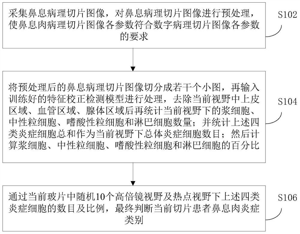
[0049] figure 1 A flow chart of a method for analyzing pathological slices of nasal polyps according to the present application is shown.

[0050] Such as figure 1 As shown, the application disclo...


PUM


Abstract

The invention discloses a nasal polyp pathological section analysis method and system and a readable storage medium, and the method comprises the steps: collecting the pathological image information of an entity section under a microscope in real time through an image collector, and transmitting the information to a processor; the processor is used for performing effective area detection on all cells on the nasal polyp pathological image, removing an epithelium area, a blood vessel area and a gland area in the current visual field, and then counting the number of plasma cells, neutrophils, eosinophils and lymphocytes in the current visual field; counting the sum of the four types of inflammatory cells as the total number of inflammatory cells in the current view; and then calculating the percentage of plasma cells, neutrophil cells, eosinophil cells and lymphocytes, finally judging the nasal polyp inflammation type of the current slice patient according to the number and proportion of the four types of inflammatory cells under the random 10 high-power lens visual fields and hot spot visual fields in the current slide, and finally storing the statistical result. The invention is helpful for individualized diagnosis and treatment and prognosis improvement of nasosinusitis patients.

Description

technical field [0001] The present invention relates to the field of pathological image processing, and more specifically, to a method, system and readable storage medium for analyzing pathological slices of nasal polyps. Background technique [0002] Nasal polyps (nasal polyps) are a common disease of the nose, which are formed due to the gradual sagging of the extremely edematous nasal and sinus mucosa under the action of gravity. Most believe that chronic infection and allergy are the possible causes of the disease. Nasal polyps are composed of highly edematous nasal mucosa, and are prone to occur on the outer wall of the nasal cavity and the top of the nose. The epithelium is pseudostratified columnar ciliated epithelium, and some epithelium metaplasia into squamous epithelium, which is caused by long-term external stimulation. Under the epithelium is edematous loose connective tissue with infiltrating inflammatory cells, including plasma cells, neutrophils, eosinophil...

Claims


Application Information

Patent Timeline
Patent Type & Authority Applications(China)
IPC IPC(8): G06T7/00G06T7/11G06V20/69G16H30/20G06N3/04G06N3/08
CPCG06T7/0012G06T7/11G06N3/084G16H30/20G06T2207/30242G06T2207/10056G06N3/045
Inventor 杨钦泰李涵生罗新邵春奎陈健宁刘子锋吴晓琦杨林黄雪琨张雅娜郑瑞吴庆武吴硕邱惠军王心悦林明珍屠佳杰
Owner 杭州迪英加科技有限公司
Why Eureka
Try Eureka