Transfection method
A bubble and cell technology, applied in the field of transfection, to achieve the effects of low-cost production, cost-effective production, and improved introduction efficiency
- Summary
- Abstract
- Description
- Claims
- Application Information
AI Technical Summary
Problems solved by technology
Method used
Image
Examples
Embodiment 1
[0185] Reference Example 1: Preparation of Ultrafine Bubble Water
[0186] Polysorbate 80 (2 g) (0.1% polysorbate 80) was dissolved in water for injection (2 L) or diluted Mclivaine buffer (pH 3.0, 2 L), and an ultrafine Bubble generator (nanoGALF TM FZ1N-02) to prepare an aqueous solution of ultrafine bubbles. Positively charged ultrafine bubbles can be generated when an acidic buffer (diluted Mclivaine buffer: pH 3.0) is used, and negatively charged ultrafine bubbles can be generated when water for injection is used.
[0187] Gases used for preparation: air (Example 3), C 3 f 8 (Example 1, 2, 4)
[0188] Bubble water flow: about 4.0L / min
[0189] Dissolving pressure: 300KPa±5%
[0190] The prepared ultra-fine bubble water was subjected to autoclaving for 30 min at 121° C.-124° C. using an autoclave as appropriate. After the sterilization, the ultrafine bubble average diameter, the ultrafine bubble density, and the d90 / d10 ratio were measured by a tracking method usin...
Embodiment 2
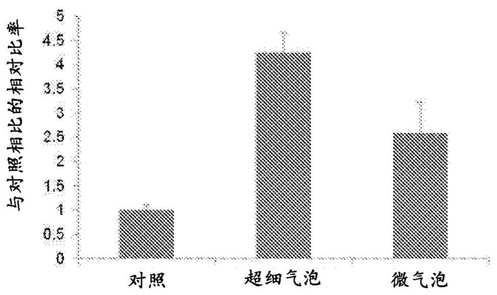
[0201] Embodiment 2 is introduced with the comparison of microbubble
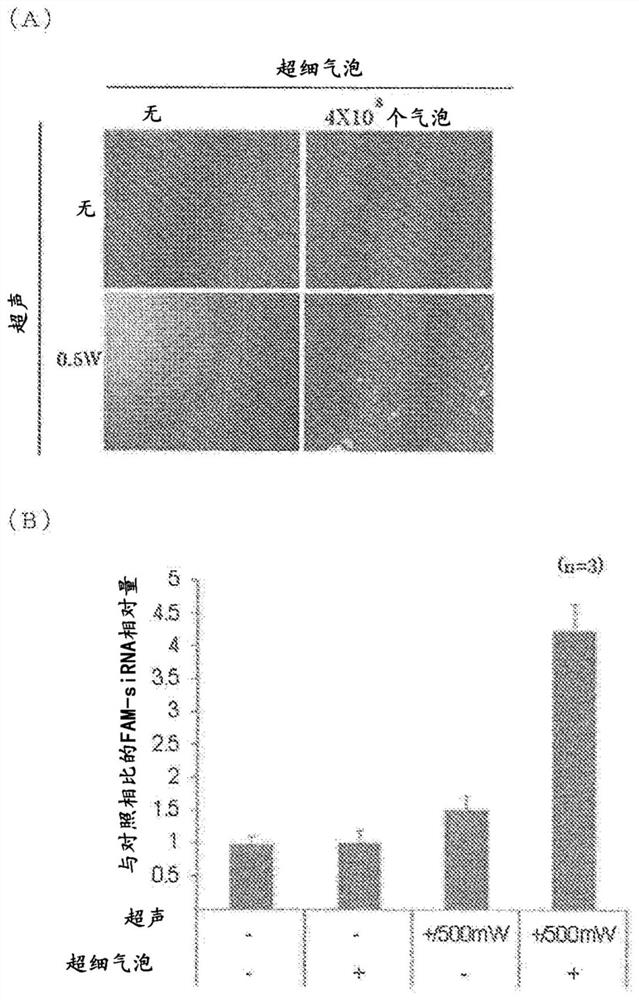
[0202] C in DMEM medium containing 10% FBS 2 C 12 cells (3×10 4 cells, 0.5 mL) were seeded in 48 wells. After removing the medium, 0.5 mL of ultrafine bubble / DMEM medium or microbubble / DMEM medium containing 5 μg of siRNA labeled with FAM fluorescence (FAM-siRNA) was added. The ultrafine bubble / DMEM medium and the microbubble / DMEM medium prepared in the following manner were used as the transfection medium: 1 L of the ultrafine bubble aqueous solution prepared in Reference Example 1 or commercially available microbubble water Add to DMEM medium powder, and add 2g / L NaHCO 3 and 10% FBS. As the microbubble water, Sonazoid (average diameter: 3 μm) containing phosphatidylserine as a phospholipid in the constituent components was used.
[0203] After adding each FAM-siRNA-containing medium, use an ultrasonic generator (NEPAGENE) at a frequency of 1 MHz and 500 mW / cm 2 The output intensity of ultrasonic ir...
Embodiment 3
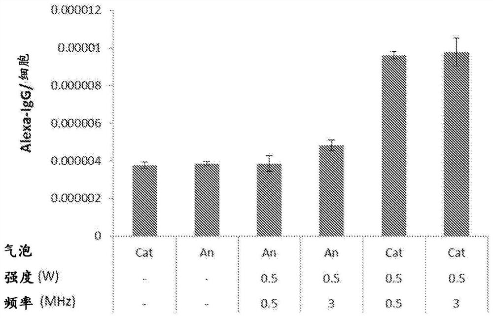
[0206] The introduction of embodiment 3 protein to skeletal muscle cell
[0207] C in DMEM medium containing 10% FBS 2 C 12 cells (6×10 4 cells, 1 mL) were seeded in 48 wells. After removing the medium, 1 mL of IgG-FITC solution diluted 100-fold with ultrafine bubble / DMEM medium was added. The ultrafine bubble / DMEM medium prepared in the following manner was used as a transfection medium: 1 L of the ultrafine bubble aqueous solution prepared in Reference Example 1 was added to the DMEM medium powder, and 2 g / L NaHCO 3 and 10% FBS.
[0208] After adding IgG-FITC-containing medium, at 500mW / cm 2 The output intensity and the frequency of 0.5MHz or 3MHz and ultrasonic irradiation using ultrasonic generator (NEPAGENE) for 10s. After ultrasonic irradiation, the cells were cultured for 2 h, then the transfection medium was removed, and the cells were cultured in DMEM medium (cultivation medium) for 48 h.
[0209] After the cultivation was completed, the fluorescence intensity ...
PUM
| Property | Measurement | Unit |
|---|---|---|
| diameter | aaaaa | aaaaa |
| diameter | aaaaa | aaaaa |
| particle size | aaaaa | aaaaa |
Abstract
Description
Claims
Application Information
 - R&D
- Intellectual Property
- Life Sciences
- Materials
- Tech Scout
- Unparalleled Data Quality
- Higher Quality Content
- 60% Fewer Hallucinations
Browse by: Latest US Patents, China's latest patents, Technical Efficacy Thesaurus, Application Domain, Technology Topic, Popular Technical Reports.
© 2025 PatSnap. All rights reserved.Legal|Privacy policy|Modern Slavery Act Transparency Statement|Sitemap|About US| Contact US: help@patsnap.com


