Fundus imaging eyepiece capable of being switched to wide angle/ultra-wide angle
An ultra-wide-angle, imaging technology, applied in ophthalmoscopes, magnifying glasses, eye testing equipment, etc., can solve problems such as signal strength reduction, optical resolution reduction, numerical aperture reduction, etc., and achieve low optical loss and high resolution. , the effect of reducing aberration
- Summary
- Abstract
- Description
- Claims
- Application Information
AI Technical Summary
Problems solved by technology
Method used
Image
Examples
Embodiment Construction
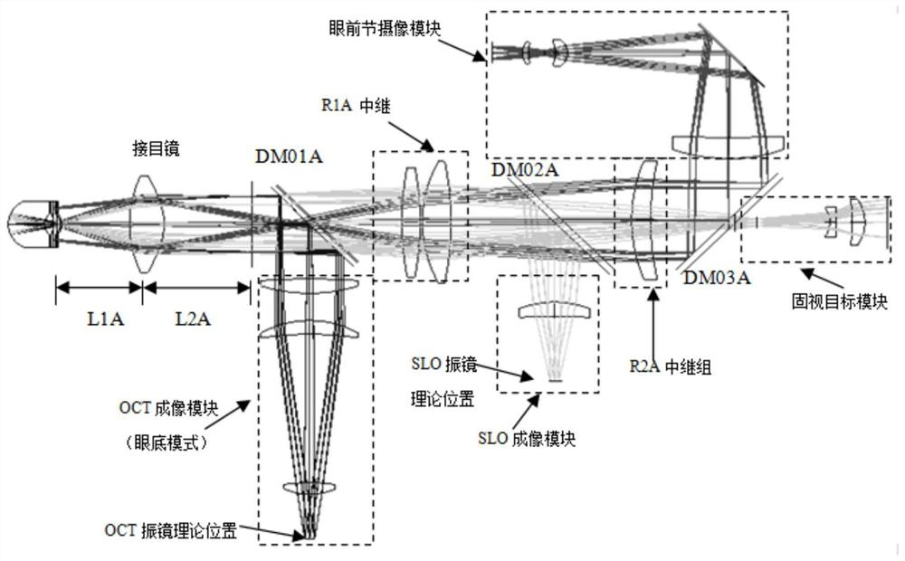
[0041] figure 1 It is an OCT ophthalmic imaging system in the prior art (Patent No. ZL2017109918972), in which the optical path is divided into four branches: OCT, cSLO, pupil camera and internal fixation lamp. Realize the switching between fundus and anterior segment image modes. There is an aspheric eyepiece at the position closest to the imaged eye in the optical path. In fundus mode, the eyepiece forms an intermediate phase plane of the fundus on its rear focal plane. OCT and cSLO pass this The fundus image is formed by two-dimensional scanning of the mesophase plane, and the eyepiece and rear optical system in the optical system can realize a 56° fundus field of view.
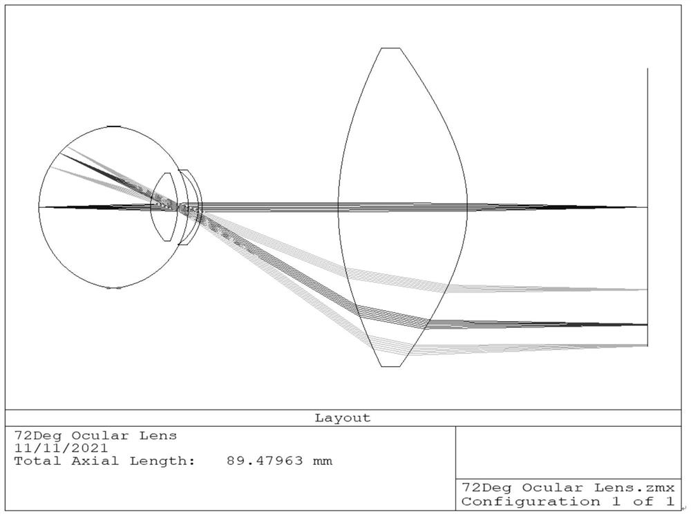
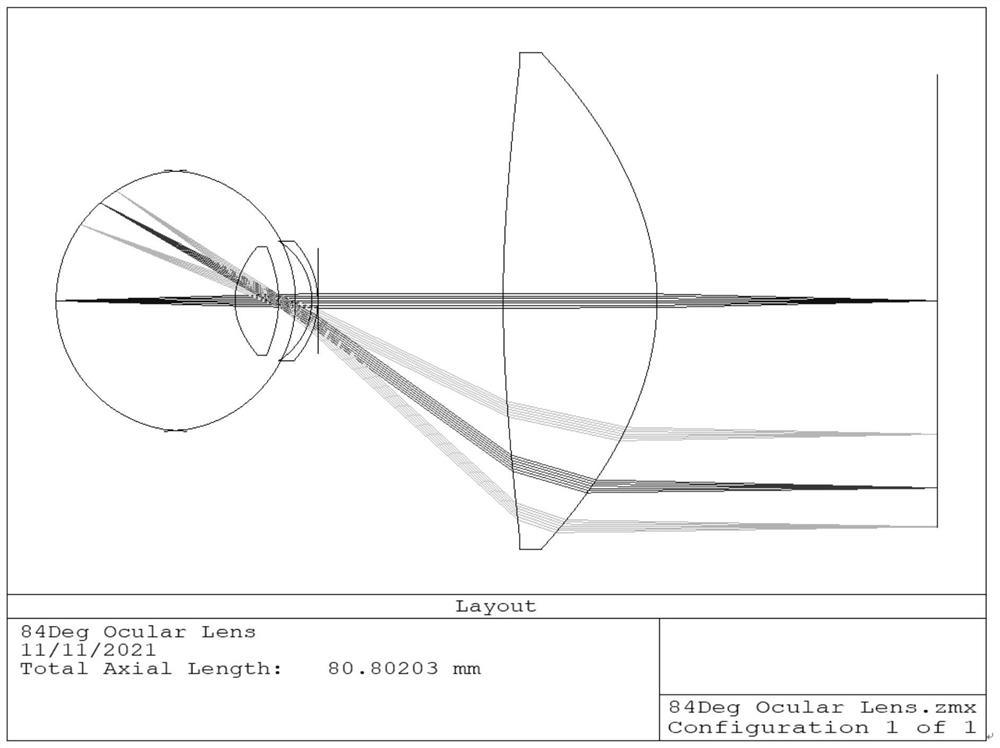
[0042] figure 2 for will figure 1 The eyepiece in the optical system is replaced by a short-focus eyepiece to achieve a larger field of view, figure 2 The eyepieces are double aspherical lenses, and the material is crown glass with medium refractive index, which realizes a field of view of 72°. imag...
PUM
 Abstract
Description
Claims
Application Information
 - R&D
- Intellectual Property
- Life Sciences
- Materials
- Tech Scout
- Unparalleled Data Quality
- Higher Quality Content
- 60% Fewer Hallucinations
Browse by: Latest US Patents, China's latest patents, Technical Efficacy Thesaurus, Application Domain, Technology Topic, Popular Technical Reports.
© 2025 PatSnap. All rights reserved.Legal|Privacy policy|Modern Slavery Act Transparency Statement|Sitemap|About US| Contact US: help@patsnap.com


