Medical image segmentation method based on boosting-unet segmentation network

A medical imaging and network technology, applied in neural learning methods, image analysis, biological neural network models, etc., can solve the problem of low image blur resistance, improve learning ability, improve performance, and reduce the number of effects

Active Publication Date: 2022-07-26
SOUTH CHINA NORMAL UNIVERSITY
View PDF7 Cites 0 Cited by
  • Summary
  • Abstract
  • Description
  • Claims
  • Application Information

AI Technical Summary

Problems solved by technology

[0005] The invention provides a medical image segmentation method based on the Boosting-Unet segmentation network, which solves the problem of low resistance to noise and image blur due to too many training parameters

Method used


Image

  • Medical image segmentation method based on boosting-unet segmentation network
  • Medical image segmentation method based on boosting-unet segmentation network
  • Medical image segmentation method based on boosting-unet segmentation network

Examples


Embodiment 1

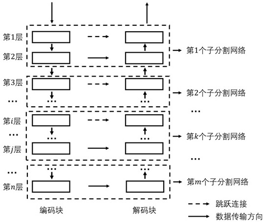
[0074] The dataset used in this example comes from a pancreatic cancer-related CT slice image dataset provided by a hospital, wherein the CT slice image dataset includes the CT slice image data of the arterial phase and the portal venous phase, and contains CT slice images of 69 cases in total. Slice images, a total of 27184 CT slice images, occupying a total of 48GB space, this example uses the constructed , layer total segmentation network, such as image 3 As shown in the figure, the segmentation of the pancreas and its pancreatic cancer in CT slice images includes the following steps:

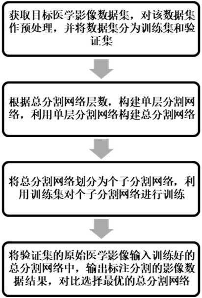
[0075] S1: Obtain the CT slice image data set, truncate and normalize the CT slice image data set: according to the grayscale interval of the CT slice image data , assuming a certain pixel value in the original image data , after truncation, the pixel value is , then after normalization, the pixel value is , perform the same operation on all CT slice image data, take the processe...

Embodiment 2

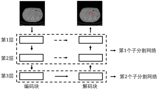
[0084] The data set used in this example is the same as that in Example 1, and this example uses the constructed of layer total segmentation network, such as Figure 4 As shown in the figure, the segmentation of the pancreas and its pancreatic cancer in CT slice images includes the following steps:

[0085] S1: Obtain the CT slice image data set, truncate and normalize the CT slice image data set: according to the grayscale interval of the CT slice image data , assuming a certain pixel value in the original image data , after truncation, the pixel value is , then after normalization, the pixel value is , perform the same operation on all CT slice image data, take the processed CT slice image data as input, and also need to back up a copy of CT slice image data as the marked and segmented CT slice image data; The annotated and segmented CT slice image dataset is divided into training set and validation set.

[0086] S2: Build contains The total segmentation networ...

Embodiment 3

[0095] The data set used in this example is the same as that in Example 1, and this example uses the constructed layer total segmentation network, such as Figure 5 As shown in the figure, the segmentation of the pancreas and its pancreatic cancer in CT slice images includes the following steps:

[0096] S1: Obtain the CT slice image data set, truncate and normalize the CT slice image data set: according to the grayscale interval of the CT slice image data , assuming a certain pixel value in the original image data , after truncation, the pixel value is , then after normalization, the pixel value is , perform the same operation on all CT slice image data, take the processed CT slice image data as input, and also need to back up a copy of CT slice image data as the marked and segmented CT slice image data; The annotated and segmented CT slice image dataset is divided into training set and validation set.

[0097] S2: Build contains The total segmentation network of ...


PUM


Abstract

The present invention provides a medical image segmentation method based on the Boosting-Unet segmentation network. The method divides the training of the total segmentation network into the training of m sub-segmentation networks. When training the k-th sub-segmentation network, the method inherits the k-1-th sub-segmentation. The convolution kernel parameters of the network greatly reduce the number of convolution kernel parameters for each training, improve the learning ability of the network and the resistance to noise and image blur. In addition, setting up multiple sub-segmentation networks improves the efficiency of the network, and realizes the The image data features are deeply extracted and the image data is finely segmented, so as to improve the overall segmentation network's ability to learn the image data features, enhance the robustness to noise disturbance information, and further improve the performance of image segmentation.

Description

technical field [0001] The invention relates to the field of medical image segmentation, in particular to a medical image segmentation method based on a Boosting-Unet segmentation network. Background technique [0002] Medical image segmentation plays a very important role in clinical diagnosis and treatment. For example, in the process of determining the location of pancreatic cancer in a patient's medical image, and dividing the edge position of pancreatic and pancreatic cancer, it is necessary to achieve accurate correlation between pancreatic and pancreatic cancer. recognition segmentation. A large number of medical images require doctors to spend a lot of time and energy to read and analyze the images, and the information obtained from CT and MRI analysis and judgment with naked eyes is very limited and uncertain. In medical imaging, because the related organs and tissues are prone to have blurred boundaries and low contrast with surrounding tissues and organs, it is e...

Claims


Application Information

Patent Timeline
Patent Type & Authority Patents(China)
IPC IPC(8): G06T7/10G06N3/04G06N3/08
CPCG06T7/10G06N3/08G06T2207/10081G06T2207/20081G06T2207/20084G06T2207/30004G06N3/048G06N3/045
Inventor 叶颀温利辉陈家炜方驰华
Owner SOUTH CHINA NORMAL UNIVERSITY
Why Eureka
Try Eureka