
Epidural and subdural hematoma detection headgear

A technology for detecting headgear and epidural, which is applied in the field of medical devices, can solve the problems of detection point data, no way to record signals, small detection range, and uncertain detection results, so as to reduce the probability of exacerbation of the disease, light weight, small size effect

Pending Publication Date: 2022-06-24
冯军峰
View PDF0 Cites 0 Cited by
  • Summary
  • Abstract
  • Description
  • Claims
  • Application Information

AI Technical Summary

Problems solved by technology

Once encountering the peak time of CT scan, there will still be a problem of waiting in line, which will bring a lot of inconvenience to the patient's family
The cost of multiple CT scans should not be underestimated, adding an invisible pressure to the patient's family
[0004] Especially in the initial stage of injury, the general patient will be sent to the hospital after being injured. During the period of sending to the hospital, there is a great demand for detection of epidural hematoma. Currently, the existing detection instruments include patch detection and Pistol-type detection, this type of detection instrument has the following problems. First, the patch-type detection range is small, and can only detect hematomas at a few points, and if you want to expand the detection range, you need to manually move the patch to change the detection position , and may also require moving the patient's head, without moving the patient's head, the patient's hematoma cannot be detected
Second, the patch type is the same as the pistol type. The detection is affected by people's subjective consciousness. The position of the hematoma is judged artificially and the size of the hematoma is artificially predicted. It can only detect the position where the hematoma is likely to appear subjectively
After arriving at the hospital, the doctor is not sure about the test results on the way, and will still ask the patient to take a CT scan. The pre-admission test did not play a substantial role
Third, whether it is a patch or a pistol-type detection instrument, it can only detect a few points at a time. There is no way to record the data and signals of the detected points. Restore the real hematoma situation of the patient

Method used


Image

  • Epidural and subdural hematoma detection headgear
  • Epidural and subdural hematoma detection headgear
  • Epidural and subdural hematoma detection headgear

Examples


Embodiment 1

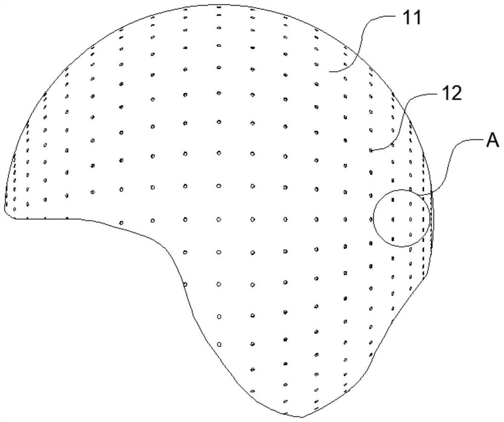
[0045] An epidural and subdural hematoma detection headgear, characterized in that it comprises,

[0046] detection components;

[0047] Wherein, the detection assembly includes a plurality of detection units, and the detection units are used to detect the thickness of the epidural hematoma at the local position of the patient's head;

[0048] Wherein, the detection component has an elastic material, and the elastic material is a headgear-like structure;

[0049] Wherein, the elastic material has a front side and a back side, the front side is the side facing the patient's head, and the back side is the side facing away from the patient's head;

[0050] The front side has the detection unit and the back side has the marking point.

[0051]At present, the mainstream methods for detecting epidural hematoma include CT detection, in addition to patch-type and pistol-type mobile detection equipment. The accuracy of CT detection is relatively high, but the CT detector is bulky an...

Embodiment 2

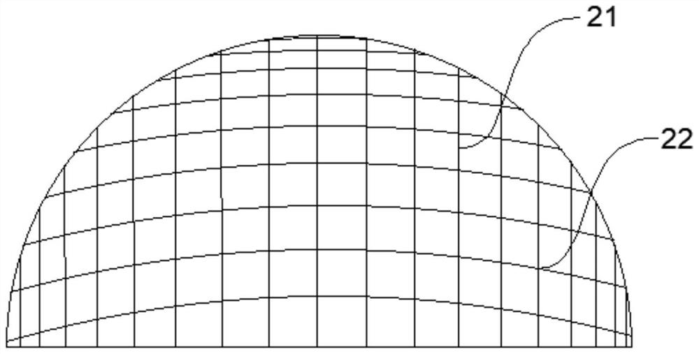
[0055] Detect auxiliary components;

[0056] Wherein, the detection auxiliary member is a mesh structure, and the mesh structure is composed of a linear structure;

[0057] Wherein, the mesh structure has no elasticity and can be bent, and the mesh structure sags naturally due to gravity;

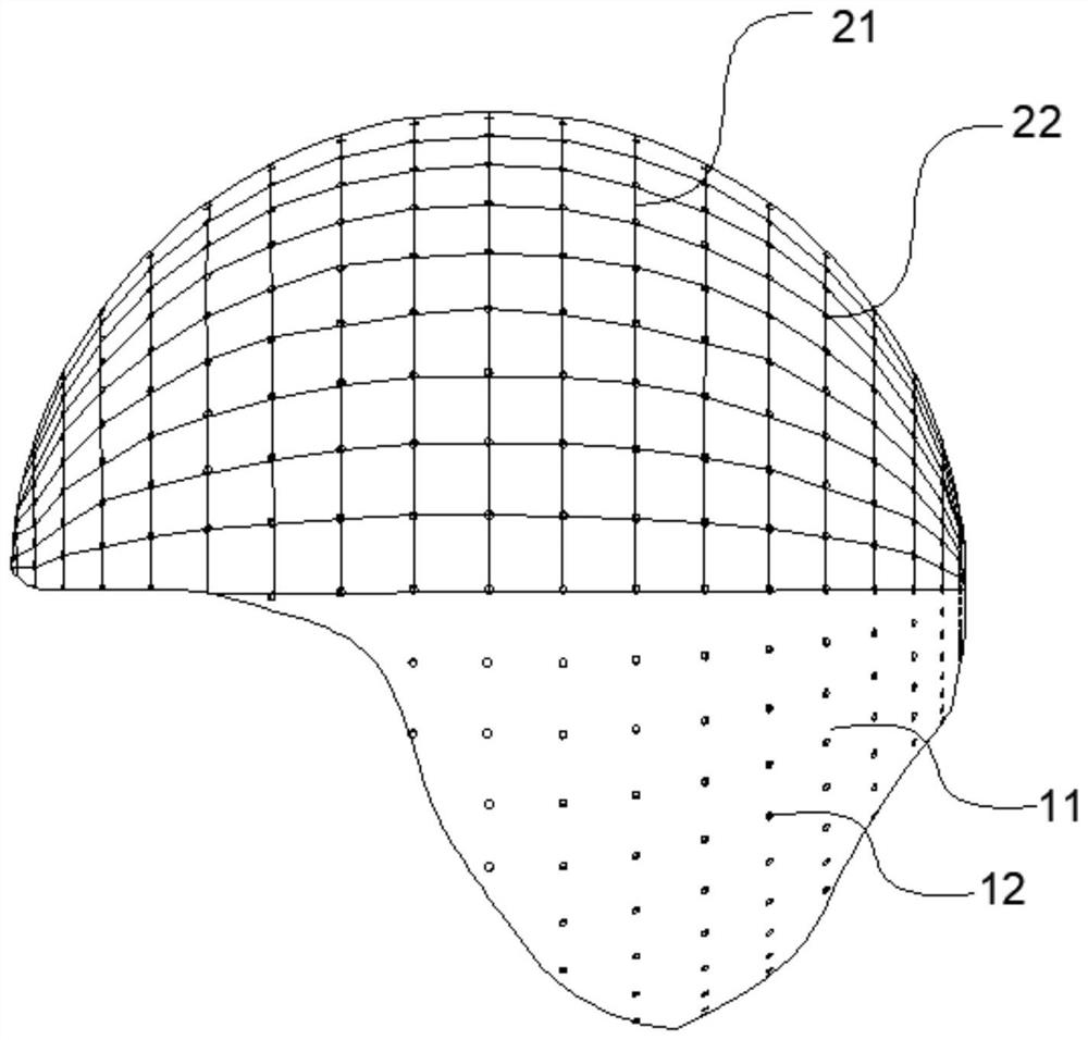
[0058] When the detection component is not affected by external force, the mesh structure can be attached to the reverse side of the elastic material;

[0059] Wherein, the intersection of the linear structure and the linear structure is a compensation reference point;

[0060] Wherein, the compensation reference point and the marked point coincide in a one-to-one correspondence.

[0061] In the actual detection situation, due to the different head shape and thickness of each patient, affected by head scars, ringworm, dandruff and other substances, there are many conditions on the skull outside the hematoma site, including the skull Relatively convex, the skull is relatively smooth, espe...

Embodiment 3

[0064] the marking point has a marking light;

[0065] Wherein, the marking lights are in the same state at the same time.

[0066] In the actual use of the headgear, with the frequent use of the headgear, the use environment includes not only the hospital, but also the place where the patient needs it such as the ambulance. In many emergency situations, doctors do not have the conditions to wear gloves for operation. One headgear can be used multiple times. The headgear is frequently picked up and put on the patient's head and removed from the patient's head. If you wear it correctly, there will be an estimated position when you hold the headgear with your hand. The contact time between the hand and the headgear is long. Lots of tiny substances stick to the marked points. When the patient wears the headgear, most of the cases will lie down and rest. The patient's head is in contact with objects such as pillows. Due to the shape of the pillow and the particularity of the str...


PUM


Abstract

The epidural and subdural hematoma detection headgear is characterized by comprising a detection assembly, a detection assembly, a detection assembly and a detection assembly, wherein the detection assembly comprises a plurality of detection units, and the detection units are used for detecting the thickness of epidural hematoma at the local position of the head of a patient; wherein the detection assembly is provided with an elastic material, and the elastic material is of a headgear-shaped structure; wherein the elastic material is provided with a front side and a back side, the front side faces the head of a patient, and the back side deviates from the head of the patient; the front side has a detection unit and the back side has mark points. Compared with a CT detector, the headgear is small in size and light in weight, and meets the use conditions of non-hospitals. A detection device is arranged on the emergency ambulance, the illness state of a patient can be monitored and recorded in real time, a doctor on the ambulance can roughly know the illness state of the patient and synchronize information to a hospital, the hospital prepares for receiving the patient in advance, and it is guaranteed that the patient can receive treatment immediately once arriving at the hospital.

Description

technical field [0001] The invention belongs to the technical field of medical devices, relates to acute epidural hematoma detection technology, in particular to an epidural and subdural hematoma detection headgear. Background technique [0002] In people's daily life, there are some unavoidable traumas, especially head injuries, which pose a great threat to human life and health. Among them, epidural hematoma is one of many threats. Epidural hematomas can occur at any age, especially in young adults aged 15-50. Epidural hematoma is a hematoma that occurs between the inner plate of the skull and the dura mater. Epidural hematoma accounts for 20-30% of traumatic intracranial hematomas; traumatic epidural hematomas often appear in a hyperacute or acute form. about 85%. Typical acute epidural hematoma is more common in young and middle-aged male patients with linear fractures, with the frontotemporal and parietotemporal areas being the most common, and the temporal area cont...

Claims


Application Information

Patent Timeline
Patent Type & Authority Applications(China)
IPC IPC(8): A61B5/107A61B5/00
CPCA61B5/1072A61B5/0075A61B5/6814
Inventor 冯军峰潘帆周至宜翁维吉
Owner 冯军峰
Learn More