
Anatomical biomechanical type 3D printing humerus greater tubercle cleavage fracture guider

A biomechanical and 3D printing technology, applied in the field of medical equipment, can solve the problems of weak fixation biomechanics, wear-through, and fixation strong protrusions to be improved, so as to increase the change of fixation angle and biomechanics, promote reset and fixation, and improve The effect of medical diagnosis and treatment level

Inactive Publication Date: 2022-07-29
THE AFFILIATED HOSPITAL OF TRADITIONAL CHINESE MEDICAL TO SOUTHWEST MEDICAL UNIV
View PDF0 Cites 0 Cited by
  • Summary
  • Abstract
  • Description
  • Claims
  • Application Information

AI Technical Summary

Problems solved by technology

[0003] For fractures of the greater tuberosity of the humerus, there are currently three major types of surgical procedures in the global clinic: cannulated screws, plates, and suture anchors. However, there is no unified gold standard for surgical methods.
Although the hollow screw is simple, practical and cheap, it also has many defects. First, for the elderly with osteoporosis, its fixation biomechanics is not strong, and it is easy to fail; second, the fixation angle is single. Since the hollow screw, the domestic The vertical fixation (90°) has always been used outside, and the fixation of the strong process needs to be improved; third, the vertical fixation is under the cartilage, and there are a series of complications after the cartilage is worn through and enters the articular surface; After reduction, there is also a lack of specialized guiding tools; fifth, there is currently no reduction or treatment tool that takes full advantage of the anatomical characteristics of the greater tubercle, and the current treatment ideas and concepts are still relatively old and not innovative

Method used


Image

  • Anatomical biomechanical type 3D printing humerus greater tubercle cleavage fracture guider
  • Anatomical biomechanical type 3D printing humerus greater tubercle cleavage fracture guider
  • Anatomical biomechanical type 3D printing humerus greater tubercle cleavage fracture guider

Examples


Embodiment 1

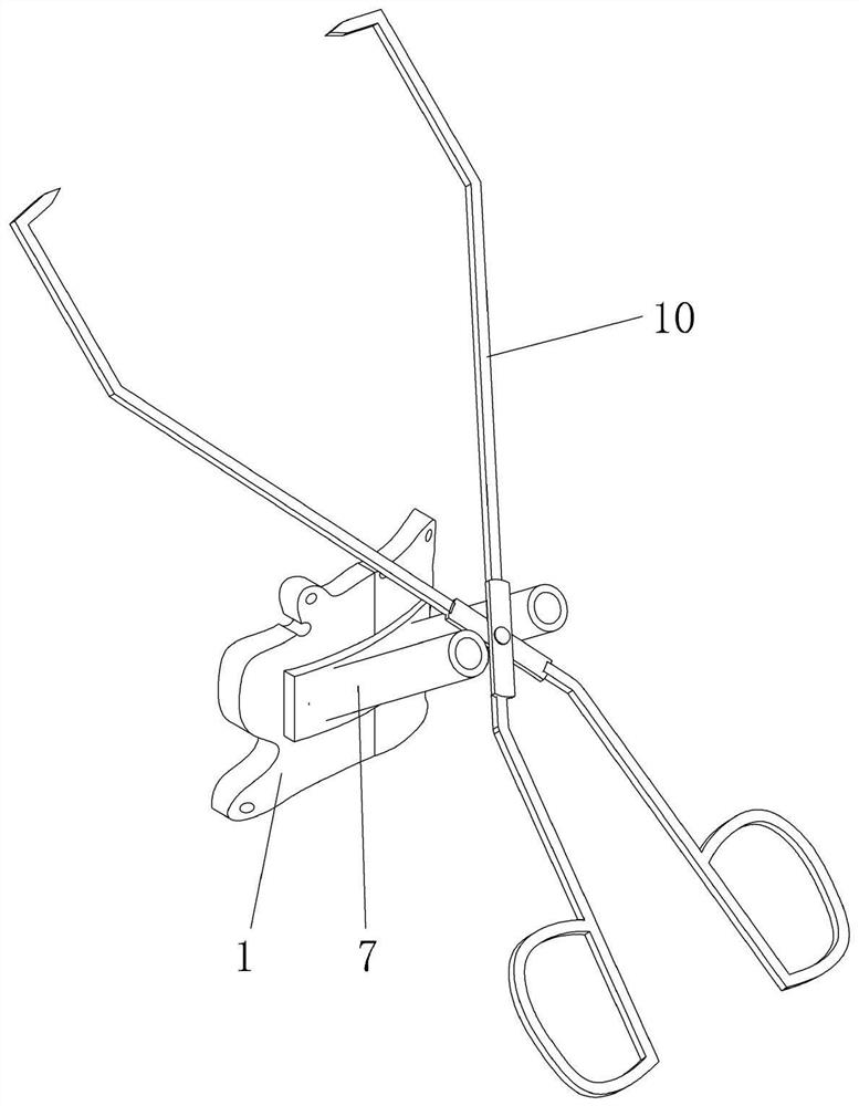
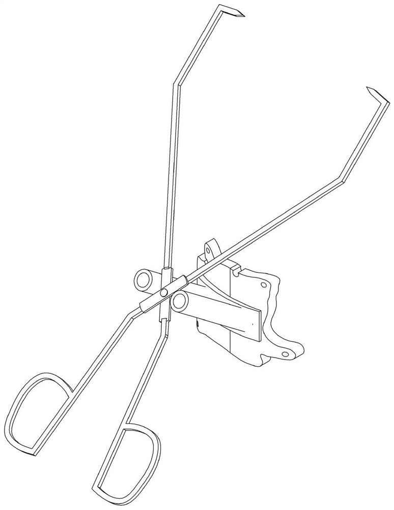
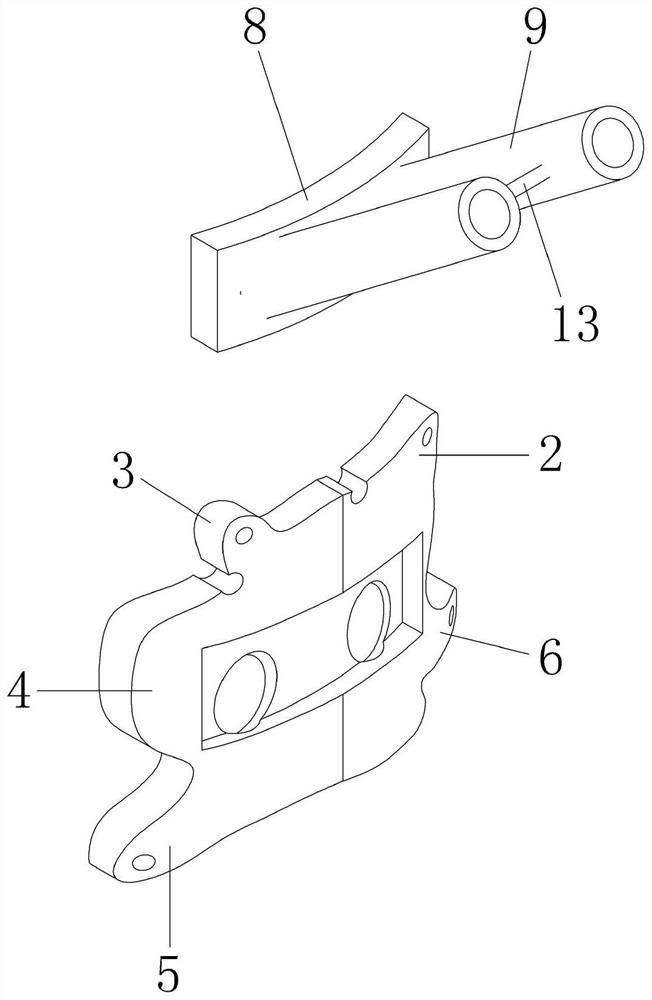
[0028] like figure 1 , figure 2 As shown, an anatomical and biomechanical 3D printed humerus greater tuberosity split fracture guide includes a guide plate 1 with an arc-like structure made based on the anatomical structure of the greater tubercle, and the guide plate 1 is integrally formed with a guide plate 1 along the circumferential direction. The first bulge 2 and the second bulge 3 made from the anatomical structure of the superior muscle, the third bulge 4 made based on the anatomical structure of the infraspinatus muscle, and the fourth bulge made based on the anatomical structure of the teres minor 5. The fifth protrusion 6 made based on the anatomical structure of the intertuberous sulcus.

[0029] like image 3 , Figure 4 As shown, a biomechanical guide sleeve portion 7 is detachably connected to the outside of the guide plate 1. The biomechanical guide sleeve portion 7 in this embodiment includes a clip 8 that is detachably connected to the guide plate 1. One...

Embodiment 2

[0034] The difference between the second embodiment and the first embodiment is that a vertical rod is welded at the lower part of the hinge part 11, and a sinker that can be adapted to the vertical rod is processed in the vertical direction in the middle of one side of the guide sleeve 9 on the card board 8. During use, the head of the towel clamp 10 clamps the acromion position, and then the vertical rod is placed in the sink cavity on the clamping plate 8 to realize the positioning of the clamping plate 8 .

[0035] The specific implementation process is as follows:

[0036]Taking Embodiment 1 as an example to illustrate, after the fracture fragment is reset, the guide plate 1 is placed on the body surface position corresponding to the greater tuberosity of the humerus, and the first protrusion 2 and the second protrusion 3 can be used to identify the supraspinatus muscle dissection. Structure, the 3rd protrusion 4 can be used to identify the infraspinatus muscle dissection...


PUM


Abstract

The invention belongs to the technical field of medical equipment, and particularly discloses an anatomical biomechanical type 3D printing humerus greater tubercle cleavage fracture guider which comprises a guide plate, a first protrusion, a second protrusion, a third protrusion, a fourth protrusion and a fifth protrusion are arranged on the guide plate in the circumferential direction, and a biomechanical guide sleeve part is detachably connected to the outer side of the guide plate. The biomechanical guide sleeve part comprises a clamping plate detachably connected with the guide plate, a plurality of guide sleeves are arranged on the side, away from the guide plate, of the clamping plate, and a plurality of first through holes communicated with the guide sleeves are formed in the clamping plate; a plurality of second through holes communicated with the first through holes are formed in the guide plate; and the fixing part is used for fixing the positions of the guide plate and the biomechanical guide sleeve part. The scheme is mainly applied to reduction and fixation treatment of the greater tuberosity cleavage fracture of the humerus, and the problems that an existing hollow screw is poor in fixation biomechanics and the like are solved.

Description

technical field [0001] The invention belongs to the technical field of medical equipment, and specifically discloses an anatomical and biomechanical 3D printed humerus greater tuberosity split fracture guide. Background technique [0002] Proximal humerus fractures, distal radius fractures, and hip fractures are also called the three most common fractures in the elderly, while the greater tuberosity fractures account for 15-20% of proximal humerus fractures, and are also a common problem that plagues the health of the elderly. disease. The greater tuberosity fracture of the humerus is not only a series of trauma diseases, it is related to the rotator cuff injury disease in the field of shoulder sports medicine, and it is also a disease frequently affected by comminuted fractures of the proximal humerus. [0003] For fractures of the greater tuberosity of the humerus, there are currently three main types of surgical procedures in the world: cannulated screws, plates, and sut...

Claims


Application Information

Patent Timeline
Patent Type & Authority Applications(China)
IPC IPC(8): A61B17/90
CPCA61B17/90
Inventor 刘刚张宝露曾胜强暴丁溯石勇郭晓光邓凯刘洋覃波罗鸿魏灵聪郝琦沈骅睿汪国友扶世杰
Owner THE AFFILIATED HOSPITAL OF TRADITIONAL CHINESE MEDICAL TO SOUTHWEST MEDICAL UNIV
Learn More