Microimaging automatic focusing image depth-of-field fusion method and related equipment

An autofocus and microscopic imaging technology, applied in the field of image processing, can solve the problems of inaccurate results, no unified judgment standard, and long time consumption

Pending Publication Date: 2022-07-29
BINGSHENG TECH WUHAN CO LTD
View PDF0 Cites 0 Cited by
  • Summary
  • Abstract
  • Description
  • Claims
  • Application Information

AI Technical Summary

Problems solved by technology

[0008] 1. In the existing technology, the subjective judgment of the operator is needed to determine whether the picture in the current field of view is clear. There is no unified judgment standard, and the judgment results of different operators may be inconsistent, resulting in unstable and inaccurate final output results
[0009] 2. In the process of adjusting the fine focus screw of the microscope, the operator needs to refocus several times to judge whether the current picture is clear, and the step size of each rotation may be different
Therefore, the clearest frames found do not have a stable gradient change, and the clearest frames may be skipped, resulting in inaccurate and time-consuming results
[0010] 3. Using artificial recognition to find pictures under different focal planes for depth fusion may not be able to find a certain part of the best picture, making the final result of depth fusion partially out of focus, which is not conducive to medical staff judging pathological conditions
[0011] 4. Manually adjust the fine focus spiral multiple times and judge the clarity of the captured frames. During long-term operation, false touches may cause the screen to move, causing the captured pictures before and after to not be in the same position.
The collected images cannot be used for depth fusion, and need to be re-acquired, with a low error tolerance rate
[0012] 5. Manual operation needs to adjust the fine focus spiral, judge the definition of the current frame, and collect pictures in sequence. In the case of multiple channels, it needs to be repeated many times.

Method used


Image

  • Microimaging automatic focusing image depth-of-field fusion method and related equipment
  • Microimaging automatic focusing image depth-of-field fusion method and related equipment
  • Microimaging automatic focusing image depth-of-field fusion method and related equipment

Examples


Embodiment Construction

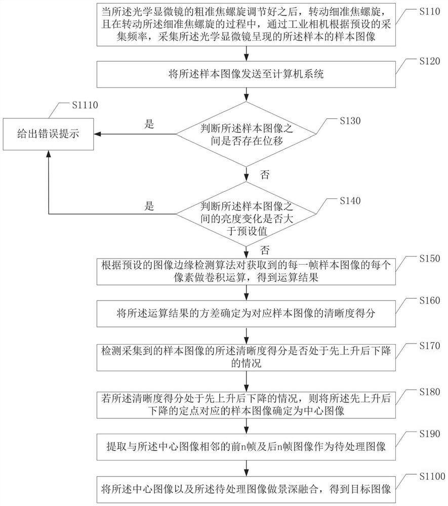
[0071] The technical solutions in the embodiments of the present invention will be clearly and completely described below with reference to the accompanying drawings in the embodiments of the present invention. Obviously, the described embodiments are part of the embodiments of the present invention, but not all of the embodiments. Based on the embodiments of the present invention, all other embodiments obtained by those of ordinary skill in the art without creative efforts shall fall within the protection scope of the present invention.

[0072] It is to be understood that, when used in this specification and the appended claims, the terms "comprising" and "comprising" indicate the presence of the described features, integers, steps, operations, elements and / or components, but do not exclude one or The presence or addition of a number of other features, integers, steps, operations, elements, components, and / or sets thereof.

[0073] It is also to be understood that the termin...


PUM


Abstract

The embodiment of the invention discloses a microscopic imaging automatic focusing image depth-of-field fusion method. The method is applied to an image depth-of-field fusion system, the system comprises a microscope system, an industrial camera and the like, and the method comprises the following steps: after a coarse focusing screw of an optical microscope is adjusted, rotating a fine focusing screw, and collecting a sample image through the industrial camera; judging whether displacement exists between the sample images and whether the brightness change is greater than a preset value through a computer system; calculating the definition score of each sample image; if displacement does not exist between the sample images, the brightness change is not greater than a preset value, and the definition scores of the sample images are firstly increased and then decreased, determining the image with the highest definition score as a central image; and extracting multiple frames of images adjacent to the central image as to-be-processed images, and performing depth-of-field fusion on the central image and the to-be-processed images to obtain a target image. According to the scheme, the definition of the target image can be improved, the sensor is used for collecting channel information, and manual participation is reduced.

Description

technical field [0001] The invention relates to the technical field of image processing, in particular to an image depth-of-field fusion method and related equipment for automatic focusing of microscopic imaging. Background technique [0002] In recent years, with the gradual unveiling of the mysteries of life, the technology of medical research has become increasingly sophisticated and complex. Microscopic imaging technology is increasingly used in the field of biomedicine. Among them, fluorescence microscopy is a very sensitive and effective technique, and almost all organic molecules can be observed and analyzed directly or after appropriate chemical treatment. In medicine, the diseased tissue and cells are marked by fluorescent staining and then photographed by a camera to obtain a clear and complete fluorescent staining map of the diseased tissue, which is provided to pathologists for post-diagnosis, which is a widely used method in current medical diagnosis. [0003]...

Claims


Application Information

Patent Timeline
Patent Type & Authority Applications(China)
IPC IPC(8): G02B21/00G02B21/24G02B21/36G06T7/13G06T7/55G06T5/50
CPCG02B21/008G02B21/242G02B21/244G02B21/367G06T7/13G06T7/55G06T5/50G06T2207/10004
Inventor 章明
Owner BINGSHENG TECH WUHAN CO LTD
Why Eureka
Try Eureka