Multi-modal cerebral apoplexy lesion segmentation method and system based on small sample learning

A multi-modal, small-sample technology, applied in neural learning methods, image analysis, biological neural network models, etc., can solve problems such as unclear boundaries, boundary restrictions, and different sizes of lesion information, to make up for insufficient data samples, The effect of improving accuracy

Pending Publication Date: 2022-07-29
SHANTOU UNIV
View PDF0 Cites 6 Cited by
  • Summary
  • Abstract
  • Description
  • Claims
  • Application Information

AI Technical Summary

Problems solved by technology

Each modality has different characteristic information, and the amount of lesion information for ischemic stroke is also different; although CT images have a wide range of applications, they can be used in various systems and parts of the body, and can find most of the lesions. However, there are certain restrictions on accurately displaying the lesion site and the boundary of the lesion site, and the boundary is not clear

Method used


Image

  • Multi-modal cerebral apoplexy lesion segmentation method and system based on small sample learning
  • Multi-modal cerebral apoplexy lesion segmentation method and system based on small sample learning
  • Multi-modal cerebral apoplexy lesion segmentation method and system based on small sample learning

Examples


Embodiment Construction

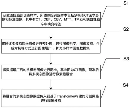
[0029] In order to make the objectives, technical solutions and advantages of the present invention clearer, the present invention will be further described in detail below with reference to the accompanying drawings.

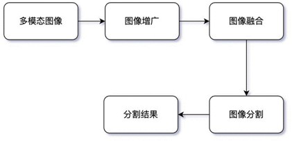
[0030] like figure 1 , figure 2 As shown in the figure, the present invention first conducts image augmentation training for multi-modal images through generative confrontation, and then extracts different feature information in multiple modalities through multi-modal fusion, and finally puts the fused image into the Transformer. The semantic segmentation network segmented the image and finally obtained the prediction result.

[0031] Firstly, the data augmentation operation is performed on the multimodal image. Image data augmentation includes image deformation, Gaussian filtering and denoising, image scaling, and generation of adversarial image augmentation.

[0032] Among them such as image 3 The specific operation of the image augmentation of the gene...


PUM


Abstract

The invention discloses a multi-modal cerebral apoplexy lesion segmentation method based on small sample learning, and the method comprises the steps: obtaining an original brain training sample which comprises a multi-modal CT medical image and an annotation image, and the original training sample comprises CT, CBF, CBV, MTT, TMax and ischemic cerebral apoplexy lesion tags; the method comprises the following steps: preprocessing a multi-modal medical image, carrying out image augmentation through modes of image deformation, image scaling, generative adversarial and the like, and expanding a small sample image data set; registering the multi-modal image after data augmentation, taking a reference image as a CT image, and performing pixel-level fusion on the multi-modal image after registration; and the fused multi-modal image data is transmitted to a segmentation network constructed based on Transform to carry out image segmentation. The invention further discloses a system using the method. According to the method, the influence on a medical image data segmentation task caused by insufficient data samples is improved, more focus image information is obtained through multi-modal image fusion, and the accuracy of image segmentation is improved.

Description

technical field [0001] The invention relates to the field of human brain CT detection, in particular to a multimodal CT image ischemic stroke lesion segmentation method and system based on Transformer deep learning network, small sample learning and multimodal analysis. Background technique [0002] Ischemic stroke is one of the most common causes of death and disability worldwide. It is caused by the occlusion of arteries in the brain, resulting in a lack of oxygen and ultimately the death of the affected brain tissue. Brain imaging plays a crucial role in the diagnosis and treatment decisions of ischemic stroke. However, the detection and evaluation of stroke lesions requires a considerable amount of time for radiologists. The following are the results of image semantic segmentation in medical imaging in recent years. [0003] In order to improve the speed and accuracy of ischemic stroke diagnosis, reliable automatic lesion segmentation methods are urgently needed. In ...

Claims


Application Information

Patent Timeline
Patent Type & Authority Applications(China)
IPC IPC(8): G06T7/00G06T7/11G06N3/04G06N3/08
CPCG06T7/0012G06T7/11G06N3/08G06T2207/10081G06T2207/20021G06T2207/20081G06T2207/20084G06T2207/30016G06N3/045
Inventor 马祥园张会凌陈盈嘉
Owner SHANTOU UNIV
Why Eureka
Try Eureka