Lattice for testing multiple biological molecule and use method thereof

A technology of biomolecules and dot arrays, applied in biological testing, measurement devices, analytical materials, etc., can solve the problems that cannot meet the needs of large-scale determination of the expression and intracellular location of different proteins

Inactive Publication Date: 2004-02-04
王颖剑
View PDF2 Cites 2 Cited by
  • Summary
  • Abstract
  • Description
  • Claims
  • Application Information

AI Technical Summary

Problems solved by technology

These methods cannot meet the needs of large-scale determination...

Method used


Image

  • Lattice for testing multiple biological molecule and use method thereof
  • Lattice for testing multiple biological molecule and use method thereof
  • Lattice for testing multiple biological molecule and use method thereof

Examples


example 1

[0074] Example 1: Making antibody arrays.

[0075] Antibody (approximately 0.5 μg / μl) was first spotted onto the nylon membrane using a mechanical spotter. The spotter drops approximately 80 nanoliters of antibody solution (approximately 40 nanograms) onto each spot using a 0.6 mm tip. The distance between points is about 0.6 mm. Antibodies are non-covalently bound to the nylon membrane. The antibody dot array prepared by this method can be used immediately or kept at 4°C within 48 hours.

example 2

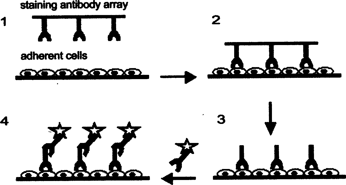
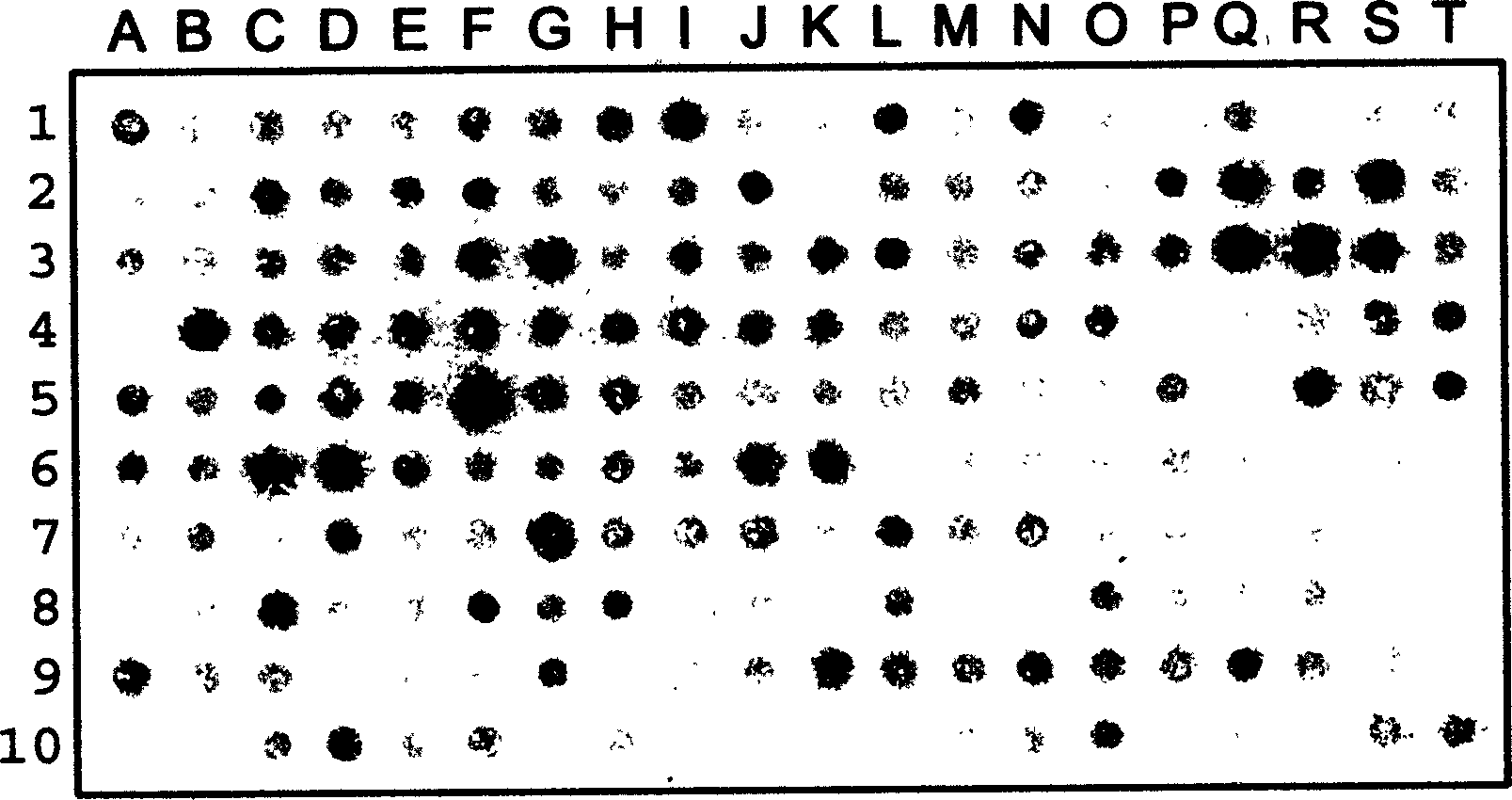
[0076] Example 2: Using antibody arrays in immunochemical staining.

[0077] This example is immunochemical staining using antibody arrays prepared by the method described in Example 1. This array contains 200 antibodies.

[0078] MDCK cells were cultured on coverslips for 2 days until confluence. The cells were then fixed and permeabilized in a methanol / acetone (1:1 ratio) mixed solution at -20°C for 10 minutes. After washing with saline, MDCK cells were exposed to the antibody array for about 1 hour. The antibody array support is then separated from the cells and their support. After the cells were washed again with normal saline, the alkaline phosphatase-labeled secondary antibody was added to react for half an hour. After washing, staining was observed by chromogenic reaction using 5-bromo-4 chloro-indolyl-phosphatase (BCIP) and nitroblue tetrazolium (NBT) as substrates. The color reaction was terminated by washing with saline. The developed image is scanned by a dig...

example 3

[0079] Example 3: Using antibody arrays in fluorescent immunochemical staining.

[0080] In this example, the spotter drops approximately 10 nanoliters of antibody solution (approximately 5 ng) onto each spot using a 0.3 mm tip. The distance between points is about 0.3mm. then apply Figure 1 Antibody arrays were used to stain A431 cells as indicated in . Similar to Example 2, except that a fluorescently labeled secondary antibody was used, and the staining was observed under a fluorescent microscope (see Figure 3). Cells were bound with fluorescently labeled secondary antibodies for half an hour. After washing, the cells were observed under a fluorescent microscope. Cells are stained by antibody at several different locations, while no diffusion of antibody is seen, as can be seen from the fact that different antigens have different subcellular sites and the areas between immobilized antibodies are not stained . Regular fluorescent spots can be observed under low magnif...


PUM


Abstract

In the present invention, a biological reagent is fixed on the first supporting body and the biological reagent fixed on the first supporting body is contacted with biological molecule fixed on the second supporting body when the dot array is used to detect the biological molecule fixed on the second supporting body. At least one biological reagent is combined with biological molecule on the second supporting body, and then the first supporting body is separated from the second one as at least one biological reagent is separated from the first supporting body to combine on the second supporting body.

Description

(1) Technical field: [0001] The present invention relates to a method for detecting biomolecules, in particular to a method of contacting a protein sample immobilized on a second support with an antibody array immobilized on a first support, and the antibodies will leave the first support A method for detecting multiple proteins by combining with a protein sample on a second support. This method can be widely used to detect the expression, activation and function of multiple proteins. (two) background technology: [0002] The rapid development of science and technology has promoted the progress of life science research, biotechnology research, drug development and research and other fields. Therefore, it is becoming more and more important to provide an efficient method for detecting proteins that are the basis of life. [0003] The emergence of a large number of biological reagents, such as tens of thousands of DNA clone sequences, numerous antibodies and recombinant prot...

Claims


Application Information

Patent Timeline
IPC IPC(8): G01N33/53G01N33/543G01N33/68
Inventor 王颖剑
Owner 王颖剑
Why Eureka
Try Eureka