Integrated image registration for cardiac nuclear magnetic resonance data perfusion

A magnetic resonance and imaging technology, which is applied in the directions of using the nuclear magnetic resonance imaging system for measurement, magnetic resonance measurement, and image data processing, and can solve problems such as difficulty in segmenting the endocardium.

Inactive Publication Date: 2004-05-12
SIEMENS AG
View PDF0 Cites 4 Cited by
  • Summary
  • Abstract
  • Description
  • Claims
  • Application Information

AI Technical Summary

Problems solved by technology

However, when there is no contrast agent in the LV, it is difficult to segment the endocardium

Method used


Image

  • Integrated image registration for cardiac nuclear magnetic resonance data perfusion
  • Integrated image registration for cardiac nuclear magnetic resonance data perfusion
  • Integrated image registration for cardiac nuclear magnetic resonance data perfusion

Examples


Embodiment Construction

[0017] Detailed Description of Preferred Embodiments

[0018] The present disclosure provides a comprehensive image registration algorithm for segmenting cardiac muscle or myocardium ("MC"). A sequence of magnetic resonance ("MR") images of the heart is acquired after contrast agent injection. Analysis of the perfusion of contrast media into the myocardium requires segmentation of the MCs in each acquired image. This segmentation task is particularly difficult due to the rapidly changing contrast in the imagery. Thus, the present disclosure provides an information registration framework that integrates two channels of information, pixel intensity and local gradient information, to reliably and accurately segment the myocardium.

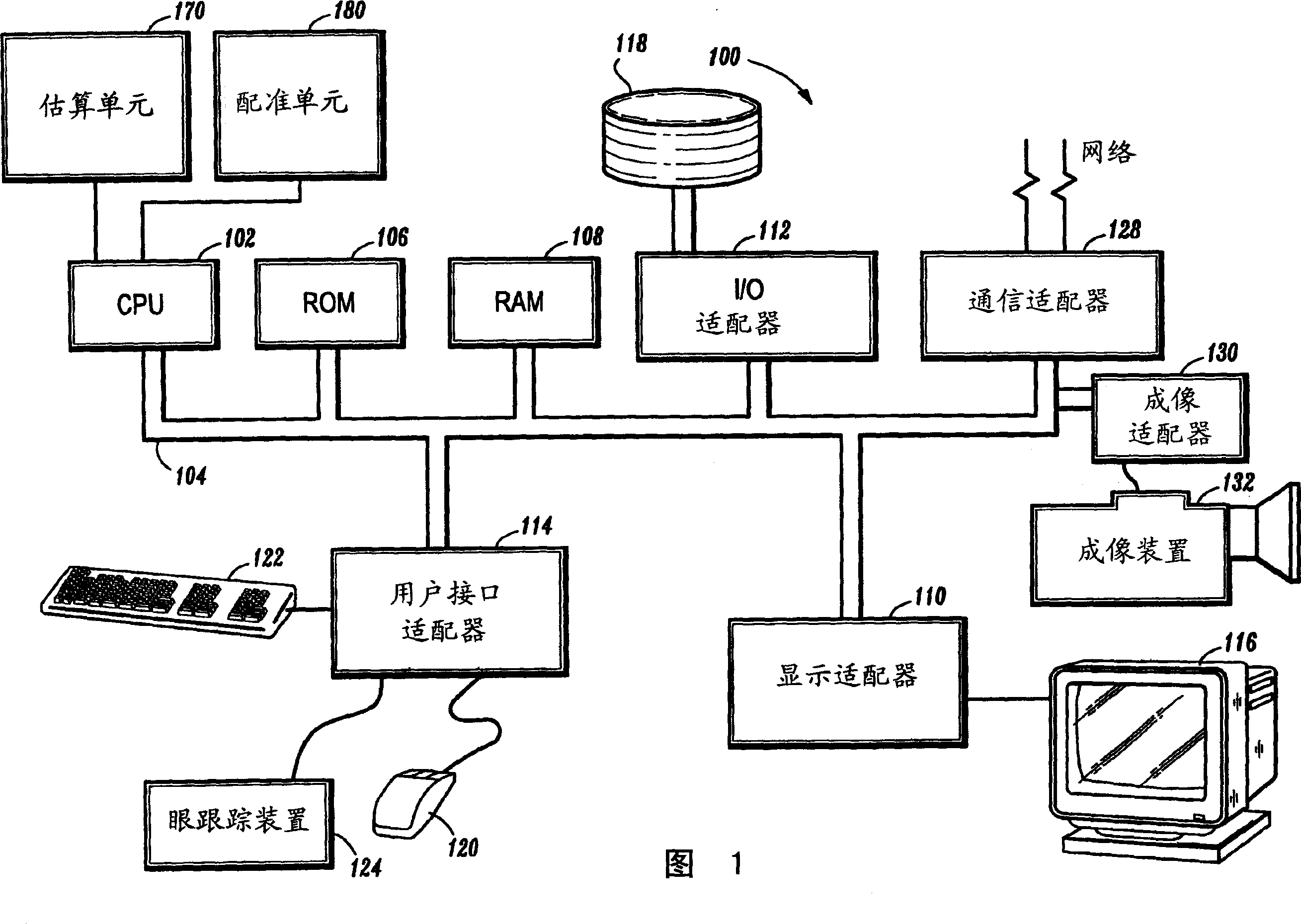
[0019] FIG. 1 shows a block diagram of a system 100 for comprehensive image registration using cardiac magnetic resonance perfusion data, in accordance with an illustrative embodiment of the disclosure. System 100 includes at least one processor or...


PUM


Abstract

A system and method for integrated image registration of cardiac magnetic resonance perfusion data are provided, where the system includes an estimation unit for estimating an edge parameter, a registration unit in signal communication with the estimation unit for registering the edge parameter in correspondence with a template-match between pixels in a first input image and pixels in a second input image, and a CPU in signal communication with said estimation unit and said registration unit for detecting a changed pixel location in accordance with said registration when the entropy of said second image is increased; and where the corresponding method for integrated image registration is of cardiac magnetic resonance perfusion data includes providing a contoured region of interest on a first image having a plurality of pixels, receiving a second image corresponding to a time other than that of said first image, calculating pixel intensities for the second image, estimating edge parameters for at least one of said first and second images, calculating a template-match between said first and second images, selecting a pair of pixel locations from at least one of said first and second images in accordance with said template-match, registering the estimated edge parameters in correspondence with said calculated template-match for a pair of pixel values at said selected locations in said first image and a pair of pixel values at said selected locations in said second image, and detecting a changed pixel location in accordance with said registration.

Description

[0001] Cross References to Related Applications [0002] This application claims priority to US Provisional Patent Application Serial No. 60 / 411,244, filed September 17, 2002, which is incorporated herein by reference. technical background [0003] In appearance-based methods for object detection and / or object recognition, segmenting imagery representing objects of interest may be complicated by dynamic motion during acquisition of a temporal sequence of imagery. An example application is the segmentation of cardiac perfusion image data. [0004] Ischemic heart disease, a disorder of blood flow to the heart, typically results from excess obesity or platelet deposits, which can narrow the veins that supply oxygenated blood to the heart. Reduced blood supply to the heart is typically expressed as reduced blood perfusion to the myocardium ("MC") heart muscle. Clinically, myocardial perfusion measurements are routinely performed with single photon emission computed tomography ("...

Claims


Application Information

Patent Timeline
IPC IPC(8): G01R33/28A61B5/055G01R33/48G01R33/54G06T1/00G06T3/00G06T7/00G06T7/60
CPCG06T7/0034G06T7/003G06T7/337G06T7/35
Inventor R·班萨尔G·芬卡-利
Owner SIEMENS AG
Why Eureka
Try Eureka