Elecrocardiographic gate control collecting image method for reducing coronary artery movement fake image

A coronary artery and image acquisition technology, applied in the field of ECG-gated image acquisition, can solve the problems of inability to diagnose coronary segments, tortuous development of coronary lumen, difficult imaging, etc., to eliminate motion artifacts and reduce variability , the effect of increased sensitivity

Inactive Publication Date: 2005-03-02
吕滨
View PDF0 Cites 3 Cited by
  • Summary
  • Abstract
  • Description
  • Claims
  • Application Information

AI Technical Summary

Problems solved by technology

[0002] Since EBCT was applied to the clinical examination of heart disease in 1984, people have always believed that the slowest point of coronary artery movement in one cardiac cycle (one heartbeat) is the mid-diastolic period, which is located near the p wave of the electrocardiogram, 70% to 80% Cardiac cycle, and 80% of the cardiac cycle of the electrocardiogram is fixedly applied to the time phase of the image acquisition, which cannot be changed in the software for EBCT computer image acqu

Method used


Image

  • Elecrocardiographic gate control collecting image method for reducing coronary artery movement fake image
  • Elecrocardiographic gate control collecting image method for reducing coronary artery movement fake image
  • Elecrocardiographic gate control collecting image method for reducing coronary artery movement fake image

Examples


Embodiment Construction

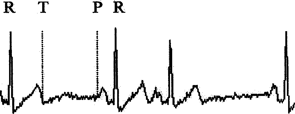
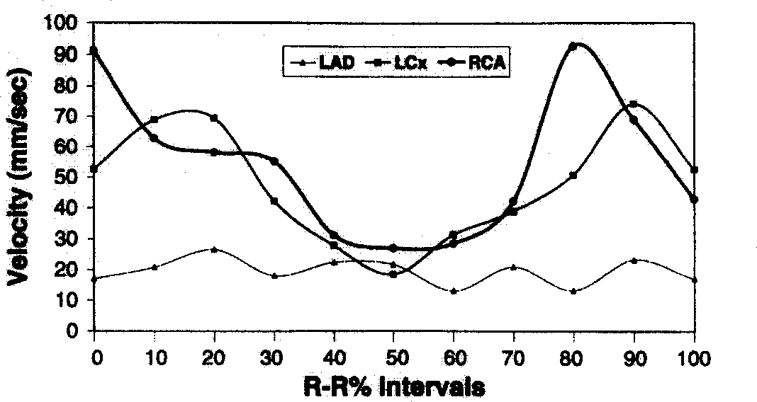
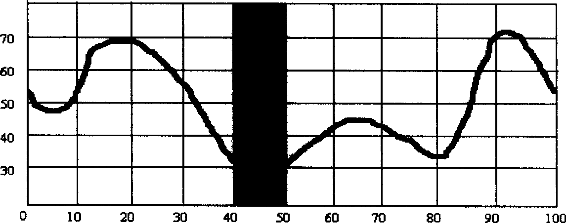
[0019] 1. EBCT cine scanning of the human heart. The EBCT scanning method is: use the cardiac cine scan mode to continuously scan each layer of the heart within one cardiac cycle. One inspection can complete the scanning of 8 different layers of the heart, and the scanning process covers the entire cardiac cycle. Cycle 0% ~ 100% RR interval; scanning layer thickness 8mm, interval between two layers 4mm. The image acquisition time of each frame is 50 milliseconds, and the interval is 6 milliseconds. The spatial position of each coronary artery is measured on each coronary artery image, so the movement distance of the coronary artery movement within the two image acquisition time (56 milliseconds) can be obtained, so the coronary artery movement velocity = movement distance ÷ time, 56 milliseconds is a fixed constant; for example figure 1 In a normal ECG, R-R indicates a cardiac cycle, T indicates T wave, that is, the end of systole, and P indicates P wave, that is, mid-diastol...


PUM


Abstract

The present invention discloses one gate controlled electrocardiographic image collecting method with reduced coronary artery motion fake image. It performs gate controlled electrocardiographic image acquisition in the lowest motion speed inside the heart beat period of the right coronary artery. The present invention obtains satisfactory image quality and has no coronary artery motion fake image radically.

Description

【Technical field】 [0001] The invention relates to an electrocardiographic gating image acquisition method for reducing coronary artery motion artifacts. 【Background technique】 [0002] Since EBCT was applied to the clinical examination of heart disease in 1984, people have always believed that the slowest point of coronary artery movement in one cardiac cycle (one heartbeat) is the mid-diastolic period, which is located near the p wave of the electrocardiogram, 70% to 80% Cardiac cycle, and 80% of the electrocardiogram cardiac cycle is fixedly applied to the time phase of the image acquisition, which cannot be changed in the software written into the EBCT computer image acquisition software. In practice, imaging is very difficult because the coronary artery moves very fast with the beating of the heart, and the lumen of the coronary artery is tortuous and small. When EBCT was used to scan the patient's heart, it was found that most patients' coronary artery images had motio...

Claims


Application Information

Patent Timeline
IPC IPC(8): A61B5/00A61B5/024A61B5/0402
Inventor 吕滨庄囡戴汝平
Owner 吕滨
Why Eureka
Try Eureka