Distributed cardiac image multi-dimensional re-building and interactive visual method

A distributed and core technology, applied in the direction of image data processing, 3D image processing, 2D image generation, etc., can solve the problems of imperfect functions, expensive, time-consuming, etc., and achieve convenient upgrades, low cost, and high efficiency Effect

Inactive Publication Date: 2005-07-27
SICHUAN UNIV
View PDF0 Cites 17 Cited by
  • Summary
  • Abstract
  • Description
  • Claims
  • Application Information

AI Technical Summary

Problems solved by technology

However, the current status of medical ultrasound imaging equipment is: expensive, single-user response, and insufficient functions
For example, the Philip 7500 real-time three-dimensional ultrasound imaging system recently launched by the Netherlands Philip Company costs more than two million yuan, but still has the disadvantage of a relatively narrow three-dimensional display range, which cannot meet the requirements of clinical diagnosis in some aspects; The 3D Cardiology Freehand system costs nearly 200,000 RMB, but in some aspects of 3D reconstruction, doctors still need to manually draw the outline of the tissue, which is not only time-consuming, but also has poor repeatability
[0003] For the diagnosis of heart disease, distributed multi-dimensional reconstruction and interactive visualization systems are very important, but there is no such system on the market, and there are no literature reports on such systems or methods

Method used


Image

  • Distributed cardiac image multi-dimensional re-building and interactive visual method
  • Distributed cardiac image multi-dimensional re-building and interactive visual method
  • Distributed cardiac image multi-dimensional re-building and interactive visual method

Examples


Embodiment Construction

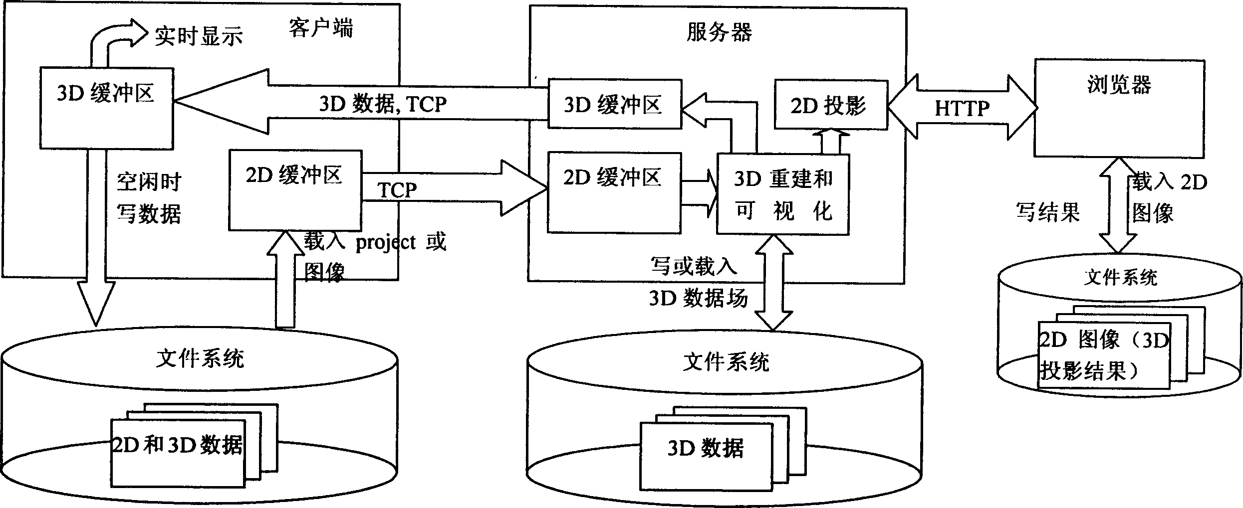
[0044] The method of the present invention requires two or more PCs with image processing capabilities, one of which is installed with server software, and the others are installed with client or / and browser software. The client / server is in CS mode, using TCP as the transmission protocol; the browser / server is in BS mode, using HTTP transmission protocol, and the image sequence is described in XML. After the server receives the XML file, it needs to convert it into an image file. After receiving the XML file, it also needs to be converted into an image file.

[0045] According to the method described in the present invention, the system structure that server, client, browser form is as follows figure 1 shown. The server accepts the request from the client or the browser, is responsible for processing the two-dimensional and three-dimensional images, and returns the processing result to the client or the browser. The client is responsible for loading the two-dimensional imag...


PUM


Abstract

A distributed method for reforming the multi-dimension heart image and interactively visualizing the reformed image includes such steps as downloading the 2D image sequence of the heart to be processed, transmitting it and processing request to server, 2D or 3D processing of said image sequence, transmitting the processed data from server to local user or remote user, and observing the resultant image via keyboard and mouse for local user or via browser for remote user.

Description

technical field [0001] The invention belongs to the field of medical image imaging and processing, in particular to a method for multi-dimensional reconstruction and interactive visualization of heart images. Background technique [0002] Cardiac ultrasound imaging is a rapidly developing field in medical imaging. Three-dimensional ultrasound can display the three-dimensional shape and anatomical structure of the heart, providing a new way for the diagnosis of heart diseases and assisting surgical operations. Therefore, ultrasonic diagnostic instruments and corresponding application software have been used clinically, and the demand for ultrasonic imaging equipment in hospitals continues to increase. However, the current status of medical ultrasound imaging equipment is: expensive, single-user response, and insufficient functions. For example, the Philip 7500 real-time three-dimensional ultrasound imaging system recently launched by the Netherl...

Claims


Application Information

Patent Timeline
Patent Type & Authority Applications(China)
IPC IPC(8): A61B5/0402A61B5/044G06T11/00G06T15/00
Inventor 杨柳饶莉郑昌琼周激流
Owner SICHUAN UNIV
Why Eureka
Try Eureka