Acquiring contrast-enhanced, T1-weighted, cine magnetic resonance images

A technology for acquiring images and magnetic resonance imaging, which is applied in the directions of magnetic resonance measurement, material analysis through resonance, and adjustment of magnetic variables. Effects of increasing equipment usage efficiency and patient throughput, increasing simplicity

Inactive Publication Date: 2005-11-09
SIEMENS MEDICAL SOLUTIONS USA INC +2
View PDF1 Cites 4 Cited by
  • Summary
  • Abstract
  • Description
  • Claims
  • Application Information

AI Technical Summary

Problems solved by technology

However, both types of cinegraphic MRI have inherently low tissue contrast, making it difficult to identify nonviable myocardium

Method used


Image

  • Acquiring contrast-enhanced, T1-weighted, cine magnetic resonance images
  • Acquiring contrast-enhanced, T1-weighted, cine magnetic resonance images
  • Acquiring contrast-enhanced, T1-weighted, cine magnetic resonance images

Examples


Embodiment Construction

[0029] The invention has too many possible implementations to be described here. Some possible implementations which are presently preferred are described below. It is important to emphasize, however, that these are descriptions of implementations of the invention, not of the invention, and that the invention is not limited to the implementations described in detail in this section, but is described in broader terms in the claims.

[0030] One implementation includes a class of pulse sequences that includes a combination of aspects of delay-enhanced magnetic resonance imaging and cinegraphic magnetic resonance imaging.

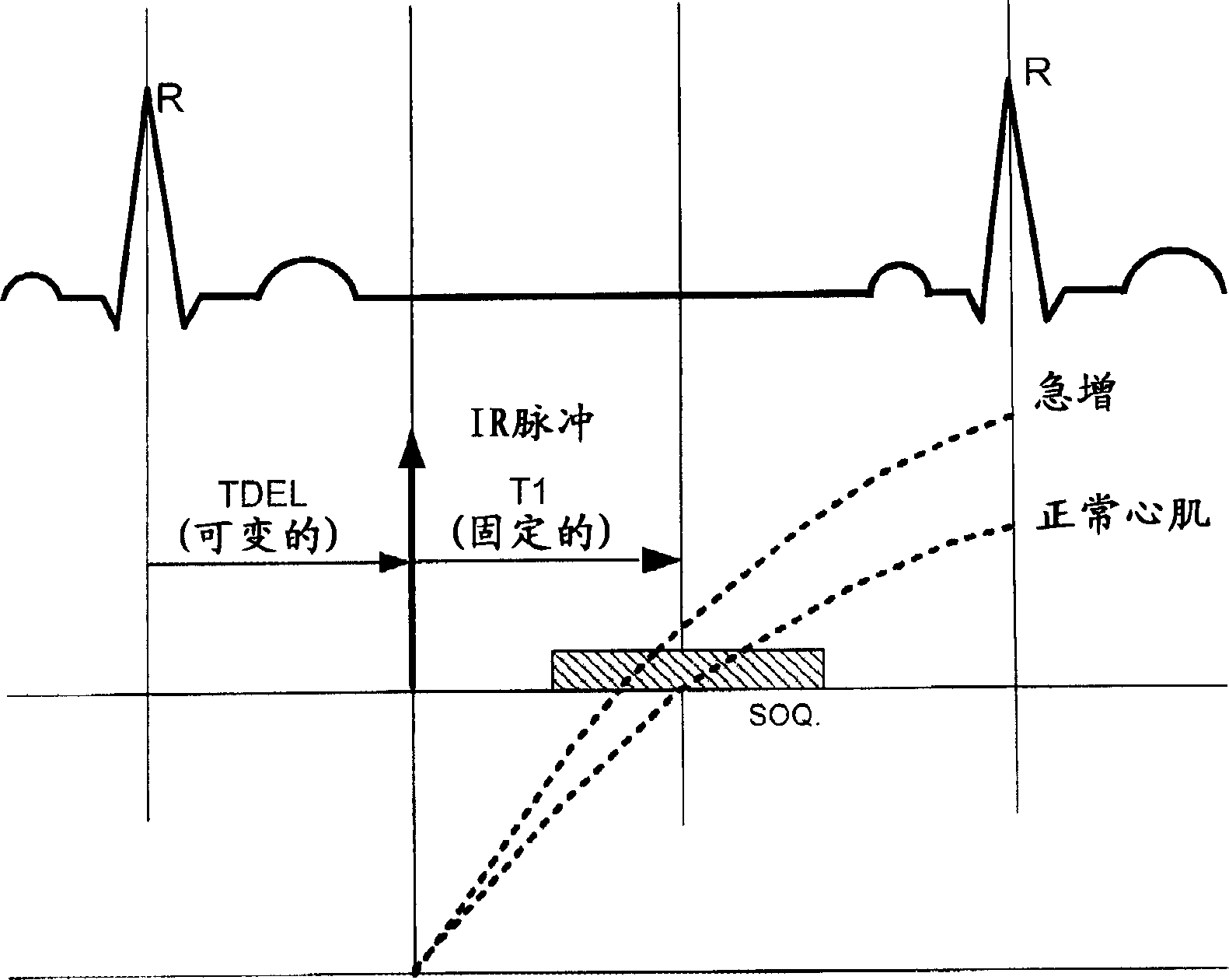
[0031] One aspect of delayed-enhanced magnetic resonance imaging is performing an inversion pulse prior to data acquisition. This inverted pulse is used to zero the normal myocardial signal to highlight hyperenhancing myocardial tissue (non-viable myocardium). Data acquisition occurs at a consistent location in the cardiac cycle, usually end-diastole.

[00...


PUM


Abstract

A method of magnetic resonance imaging of an anatomical site subject to a motion cycle (e.g., the heart during a cardiac cycle), comprising: administering a magnetic resonance contrast agent; waiting for a period of time until the contrast agent is effective to cause a selected portion of the anatomical site (e.g., dead heart tissue) has a different T1 recovery rate than other parts (normal heart tissue); give multiple time-spaced inversion recovery pulses; follow inversion recovery pulses in time spaced by known time intervals Acquire image data at data acquisition time; vary the time at which an inversion recovery pulse is given within a motion cycle so that the relevant data acquisition time is at multiple phases of the motion cycle; process image data acquired at a certain phase of the motion cycle to produce an at least a portion of the image frames of the phase; and performing the processing on a plurality of phases of the motion cycle to generate a plurality of image frames corresponding to the plurality of phases.

Description

technical field [0001] The present invention relates to magnetic resonance (MR) imaging, and more particularly, the present invention relates to magnetic resonance imaging of the heart. Background technique [0002] Identification of viable myocardium is an important, common clinical purpose, for example, in assessing the potential for revascularization therapy in patients with chronic ischemic disease (Thornhill et al., "The assessment of myocardial viability: a review of current diagnostic imaging approaches, "J. Cardiovasc. Magn. Reson. 4:381-410 (2002)). Viability imaging refers to imaging techniques used to identify abnormal myocardium (viable myocardium) that may recover function after multiple perfusions. Magnetic resonance imaging (MRI) is increasingly used to identify viable myocardium (Sandstede, "Assessment of myocardial viability by MR imaging," Eur. Radiol. 13:52-61 (2003)). Standard MRI assessment is based on the comparison of separately acquired images of th...

Claims


Application Information

Patent Timeline
Patent Type & Authority Applications(China)
IPC IPC(8): A61B5/05A61B5/055G01R33/28G01R33/567
CPCG01R33/281A61B5/055A61B5/7289G01R33/5673B42D15/00B42F7/02
Inventor J·K·金R·勒夫勒K·K·W·陈R·M·塞特塞尔A·E·斯蒂尔曼R·D·怀特
Owner SIEMENS MEDICAL SOLUTIONS USA INC
Why Eureka
Try Eureka