Liver-cancer stem cell, its separation and use
A liver cancer stem cell and separation method technology, applied in the direction of tumor/cancer cells, animal cells, vertebrate cells, etc., can solve problems such as metastasis, tumor recurrence, tumor mass shrinkage, etc.
- Summary
- Abstract
- Description
- Claims
- Application Information
AI Technical Summary
Problems solved by technology
Method used
Image

Examples
Embodiment 1
[0058] Isolation and identification of cancer stem cells in liver cancer cell lines
[0059] The specific AC133 / CD133 / 1 monoclonal antibody and magnetic bead sorting system enrich and separate stem cells from liver cancer cell lines:
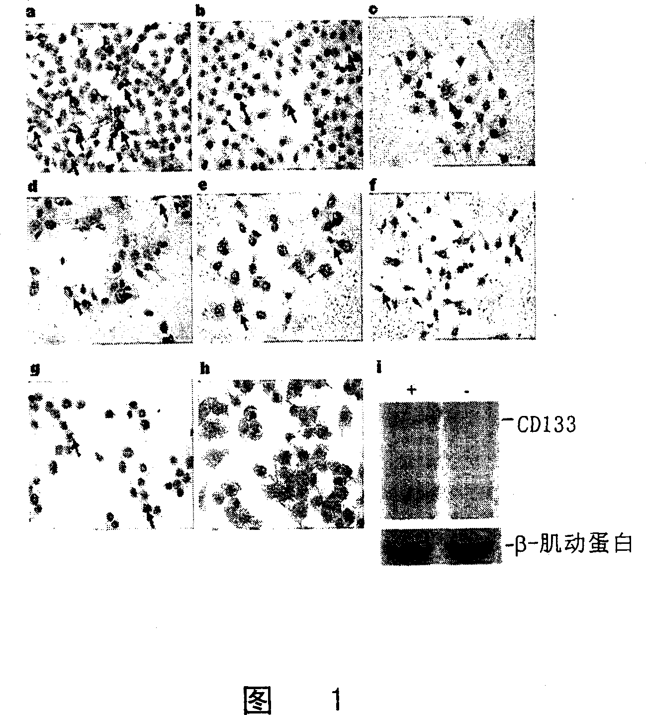
[0060] (1) Expression of stem cell marker CD133 in liver cancer cell lines: Figures 1a-f are human liver cancer cell lines, including (a) SMMC7721, (b) BEL7402, (c) Huh-7, (d) Hep3B, (e) MHCC-LM3, (f) MHCC-97L, immunocytochemical test results show that there are a small number of CD133+ cells in the above cell lines (arrow);
[0061] (2) CD133 efficiently enriched by magnetic bead sorting + Cells, of which (g) pre-sorting cells CD133 + The proportion of positive cells is low, (h) CD133 after sorting + The positive rate is greatly increased .
[0062] Western blot (i) results also confirmed this point, where "+" means CD133 after sorting + Are positive cells, "-" means CD133 after sorting - It is a negative cell, and β-actin is an internal control.
Embodiment 2
[0064] Identification and identification of cancer stem cells in human liver cancer-related tissues
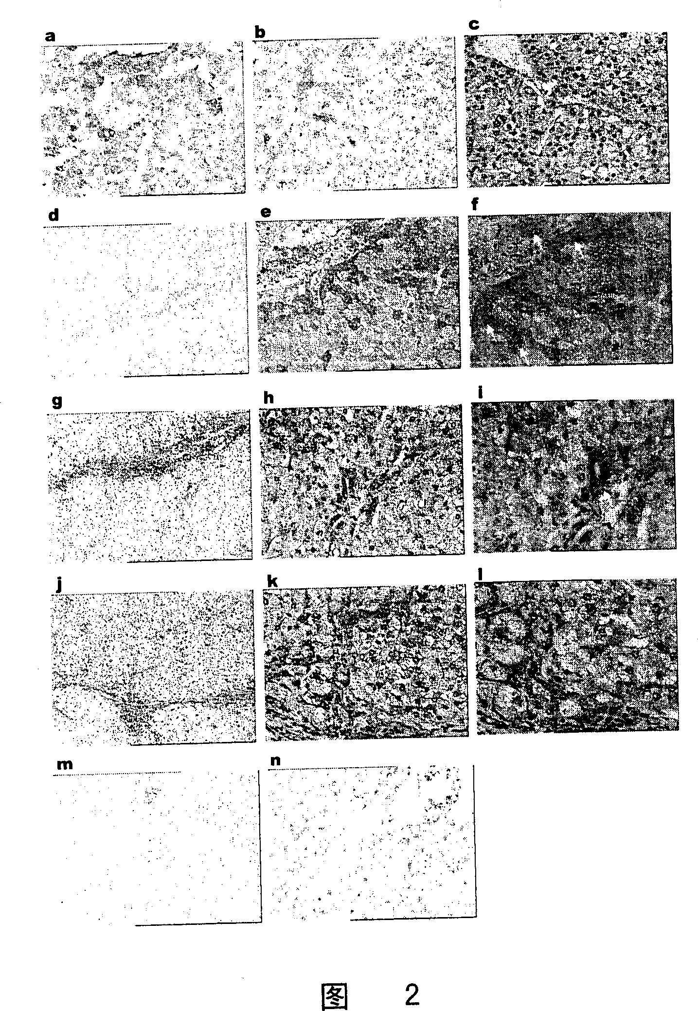
[0065] The expression of stem cell marker CD133 in paraffin-embedded liver cancer, adjacent liver, liver cirrhosis, and normal liver tissue sections (immunohistochemical detection): Figure 2a-c is liver cancer tissue, di is adjacent liver tissue, jl is liver cirrhosis tissue , M, n are normal liver tissues.
[0066] It can be seen from Figure 2 that in addition to normal liver tissues, there are few CD133-positive cells (yellow-brown) in liver cancer, para-cancerous liver, and cirrhosis tissues, and the white arrow indicates the Hering’s canal-like structure.
Embodiment 3
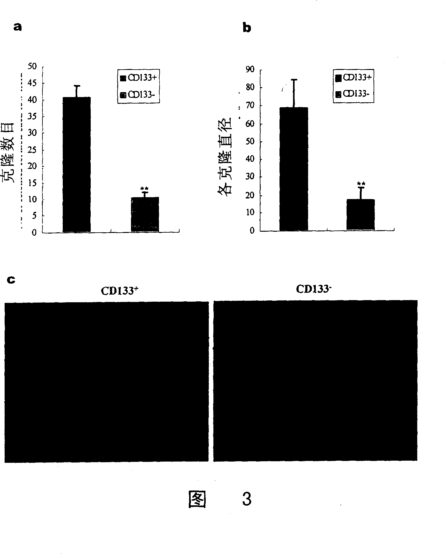
[0068] CD133 + In vitro cloning ability detection of liver cancer stem cells
[0069] CD133 was detected by soft agar colony formation assay + The cloning ability of liver cancer stem cells in soft agar, the specific steps are as follows:
[0070] (1) Pave the 6-well cell culture plate (bottom layer) with 0.6% low melting agar-stem cell culture medium containing 10% fetal bovine serum prepared at 42°C;
[0071] (2) After curing at 37℃, use 5×10 3 CD133 + Or CD133 - SMMC7721-GFP single cell-soft agar suspension covers the bottom glue;
[0072] (3) Solidify at 37°C, add 2ml of stem cell culture medium to each well, and change the fresh medium every 5 days;
[0073] (4) Three weeks later, clones with more than 15 cells were counted and photographed under an inverted fluorescence microscope, and the data was statistically analyzed.
[0074] The results are shown in Figure 3: The bar graph (a) shows the number of colonies formed, expressed as the average number ± sd (n = 12 low power f...
PUM
 Abstract
Description
Claims
Application Information
 - R&D
- Intellectual Property
- Life Sciences
- Materials
- Tech Scout
- Unparalleled Data Quality
- Higher Quality Content
- 60% Fewer Hallucinations
Browse by: Latest US Patents, China's latest patents, Technical Efficacy Thesaurus, Application Domain, Technology Topic, Popular Technical Reports.
© 2025 PatSnap. All rights reserved.Legal|Privacy policy|Modern Slavery Act Transparency Statement|Sitemap|About US| Contact US: help@patsnap.com