Radiation tomographic imaging apparatus and radiation tomographic imaging method

A tomography and imaging device technology, applied in computer tomography scanners, instruments for radiological diagnosis, diagnosis, etc., can solve the problem of high radiation exposure dose, reduce radiation exposure dose, increase efficiency, and improve operability. Effect

Inactive Publication Date: 2007-07-04
GE MEDICAL SYST GLOBAL TECH CO LLC
View PDF1 Cites 7 Cited by
  • Summary
  • Abstract
  • Description
  • Claims
  • Application Information

AI Technical Summary

Problems solved by technology

However, in the retrospective ECG method, the radiation exposure dose to the subject is higher due to the sequential projection of X-rays

Method used


Image

  • Radiation tomographic imaging apparatus and radiation tomographic imaging method
  • Radiation tomographic imaging apparatus and radiation tomographic imaging method
  • Radiation tomographic imaging apparatus and radiation tomographic imaging method

Examples


Embodiment Construction

[0031] Overview of X-ray CT device

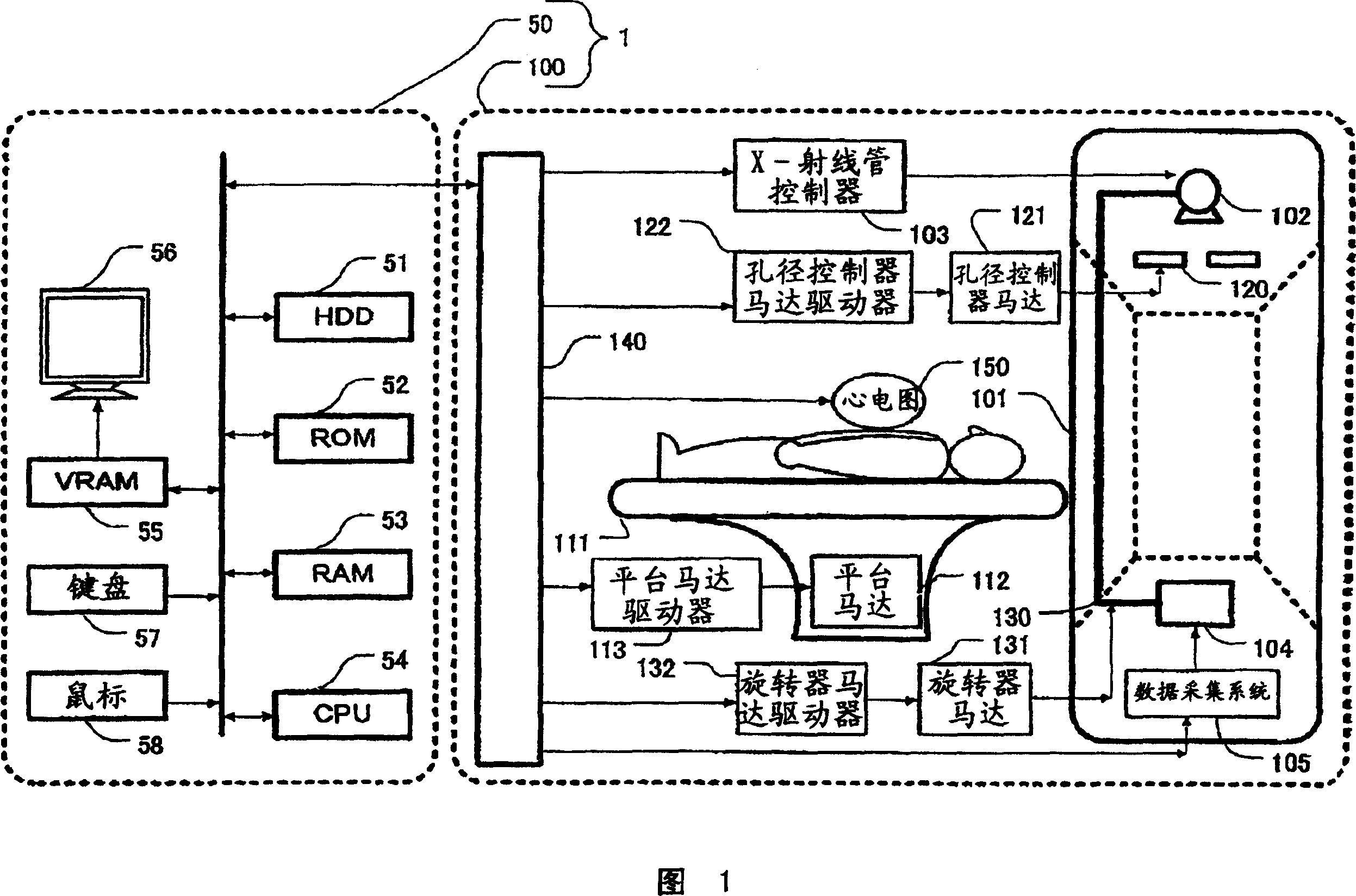
[0032] Referring now to FIG. 1 , an overview of an X-ray CT apparatus according to a preferred embodiment is shown. As shown, the apparatus includes a gantry 100 for emitting X-rays to an object and for detecting X-rays transmitted through the object, and for reconstructing X-ray tomography from data transmitted from the gantry 100 Image and operating console 50 for output and display.

[0033] The rack 100 includes a CT control unit 140 for managing entities, and is connected to various devices which will be described below.

[0034] In the frame 100, an X-ray tube 102 as an X-ray source, an X-ray tube controller 103 connected to the X-ray tube 102, a collimator with an aperture for limiting the X-ray radiation range are equipped. An instrument 120, an aperture controller motor 121 for adjusting the aperture width of the collimator 120, and an aperture controller motor driver 122 for driving the aperture controller motor 121. The X-rays...


PUM


Abstract

There is provided a radiation tomographic imaging apparatus 100 and a radiation tomographic imaging method 200 which allows the ECG information and the X-ray tube current value to be monitored while obtaining required projection data for the image reconstruction. The radiation tomographic imaging method 200 for forming a tomographic image of a subject by means of the radiation from the radiation source 102 comprises the ECG wave output step S204 for measuring the heartbeat of the heart of the subject to output as the ECG wave signal R; a variable output step S205 for varying the radiation output based on the ECG wave signal; a determining step S209 for determining the tomographic image reconstructed based on the projection data obtained by the radiation output having been varied is good or no good; and a displaying step S210 for displaying the ECG wave signal, the radiation output, and the reconstruction data area for forming a tomographic image if the image is no good.

Description

technical field [0001] The present invention relates to a radiation tomographic imaging method for performing tomographic imaging of a subject by emitting radiation from the surroundings of the subject, ie, a patient, to obtain data and process the data. More specifically, the invention relates to the reconstruction of images of cardiac regions. Background technique [0002] As a diagnostic device for damage in a subject, a radiation tomographic imaging device for acquiring a tomographic image of a subject, such as an X-ray CT device, is widely used for diagnosis. This X-ray CT apparatus is also widely used in the imaging of the cardiac region. [0003] When imaging the heart region, since the heart is always beating, the patient attaches an EKG to it to monitor the state of heart function such as systole and diastole while emitting radiation. A reconstruction method of a cardiac image is called an ECG (electrocardiogram) reconstruction method, which includes a prospective...

Claims


Application Information

Patent Timeline
Patent Type & Authority Applications(China)
IPC IPC(8): A61B6/03A61B5/0402A61B5/024
CPCA61B6/463A61B6/488A61B5/0402A61B6/503A61B6/541A61B6/032A61B5/318A61B6/03
Inventor 濑户胜
Owner GE MEDICAL SYST GLOBAL TECH CO LLC
Why Eureka
Try Eureka