
Apparatuses and methods for electrophysiological signal delivery and recording during MRI

a technology of electrophysiological signals and apparatus, applied in the field of electrophysiological signal delivery and recording during magnetic resonance imaging, can solve the problems of not providing the spatial resolution used to determine the exact location of the recorded activity in the brain, no brain imaging technology that can provide the resolution needed to study this brain activity, and several safety problems, so as to achieve safe use in high magnetic fields, reduce the effect of sar and temperature increas

Active Publication Date: 2019-06-25
THE GENERAL HOSPITAL CORP
View PDF17 Cites 0 Cited by
  • Summary
  • Abstract
  • Description
  • Claims
  • Application Information

AI Technical Summary

Benefits of technology

[0024]Another object of the present invention is to provide methods, systems and arrangements that enable a reduction of the SAR exposure while increasing signal quality and improving patient safety.
[0034]Advantageously, the exemplary embodiments of methods, systems and arrangements of the present invention can achieve lower SAR and lower temperature increase as compared to metallic or carbon fiber electrodes and leads, with approximately the same input power. The exemplary embodiment of the sensor system of the present invention may be safely used in high magnetic fields because of the minimal or no use of metals and the very weak interaction with the RF fields generated by the MRI scanner. Additionally, the multi-layered leads of this system may be less susceptible to induced ballistocardiogram currents and the sensors are less susceptible to Eddy currents that can introduce sensor heating and diminish image quality. The resistivity of the leads can also greatly increase the signal-to-noise ratio (“SNR”) of the EEG recordings.

Problems solved by technology

While the brain waves provide useful temporal information regarding the electrical activity of the brain, they do not provide the spatial resolution used for determining the exact location of the recorded activity in the brain.
Currently, there is no single brain imaging technology that can provide the resolution needed to study this brain activity.
The combination of these two conventional technologies, however, may provide several safety problems.
One such problem involves the integrity of the measurements, as the changing magnetic and RF fields of an MRI / fMRI recording can introduce undesirable artifacts into the EEG recordings.
When EEG leads are placed inside an MRI scanner, the rapidly changing RF fields may introduce voltages that obscure the EEG signals.
Further, the presence of magnetic materials within the EEG electrodes placed inside the MRI scanner and the electromagnetic radiation emitted by the EEG machine can disturb the homogeneity of the magnetic field, and possibly compromise the quality of the MRI image scans.
The introduction of the EEG equipment into the pulsed RF fields created by the MRI scanner can also present a safety hazard, especially at high static B0 fields because of Specific Absorption Rate (“SAR”) considerations.
The use of metallic electrodes and leads may cause an undesirable increase in local and whole-head SAR values, reflected in the heating of patient's tissue.
Such heating may possibly result in bodily injury to the patient, including burns, electric shock, etc.
Specifically, noise may be introduced by motion within the MRI environment during the recording of the EEG signals.
However, carbon fibers have a high and fixed conductivity that may not be easily reduced in a cost-effective and commercially feasible manner.
This is because the EEG leads can interfere with the RF fields within the MRI scanner by de-tuning the coils used in the MRI scanner, thereby resulting in a global attenuation of the received RF signal.
While these exemplary measures have assisted in improving the quality of both the EEG and MRI test results, certain problems still exist with such conventional approaches.
In particular, there are no methods, systems or arrangements that enable a concurrent recording of that EEG signals and MRI scans, which can simultaneously take into consideration the metallic nature of the electrodes and leads that results in high SAR and RF interferences, the motion and artifact noise within the MRI environment, and the patient safety in general.

Method used


Image

  • Apparatuses and methods for electrophysiological signal delivery and recording during MRI
  • Apparatuses and methods for electrophysiological signal delivery and recording during MRI
  • Apparatuses and methods for electrophysiological signal delivery and recording during MRI

Examples


Embodiment Construction

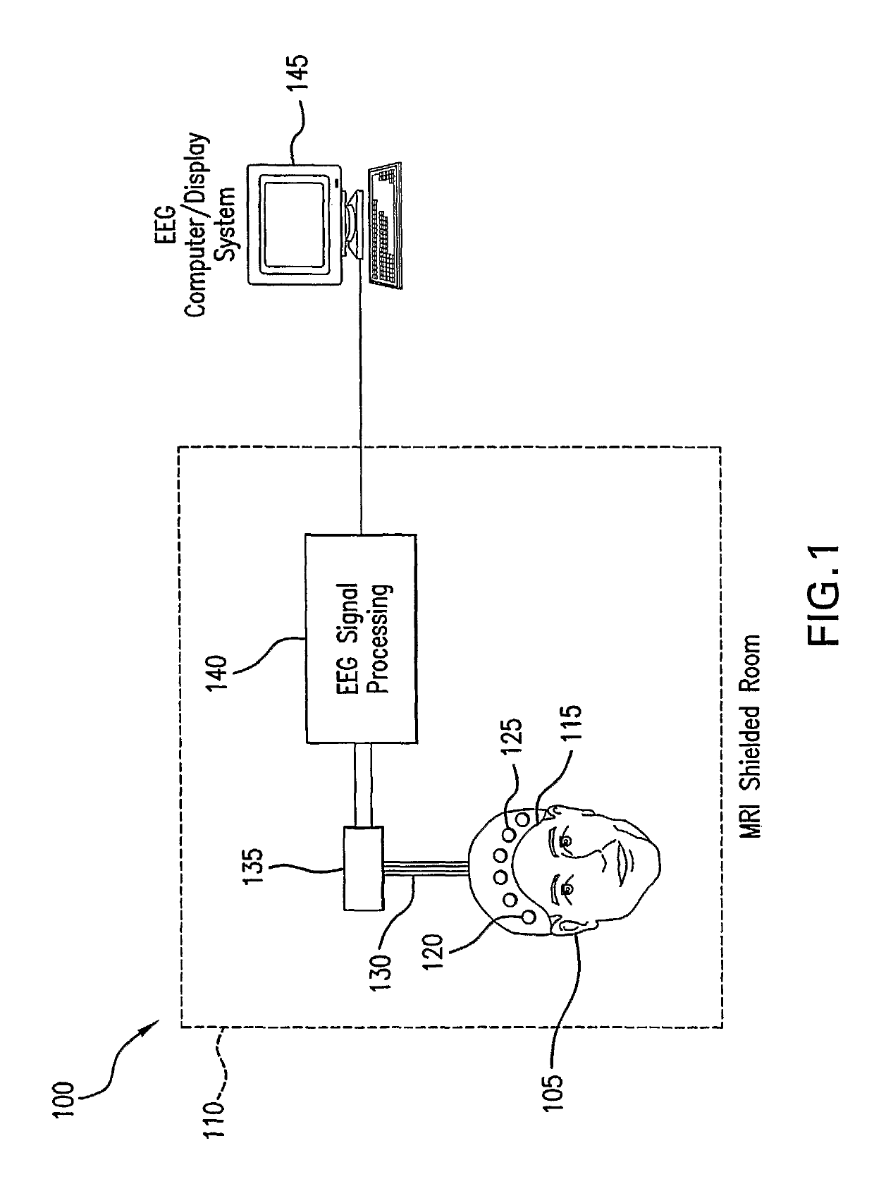
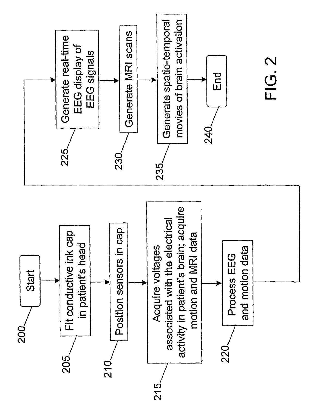
[0060]Generally, an exemplary embodiment of the present invention provides methods, systems and arrangements for the delivery and recording of electrophysiological brain signals during MRI. As used herein, electrophysiological brain signals generally refer to signals that record the electrical activity of the brain. These signals may be acquired by an electroencephalogram (“EEG”) machine, e.g., a system that can measure an electrical activity in the brain via a multitude of electrodes coupled to a patient's scalp via a cap, glue, or paste, and electrically coupled to the EEG machine via leads. Signals acquired by the EEG machine may be interchangeably referred to herein as electrophysiological brain signals, EEG signals, or brain waves.

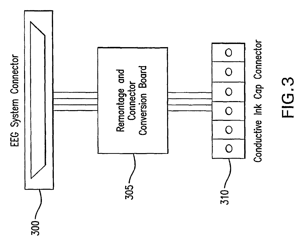
[0061]Electrodes as used herein may preferably include a conductive medium used to record an electrical signal from a patient. A group of electrodes may be attached to a cap which can fit the patient's head so as to acquire the EEG signals in accordan...


PUM


Abstract

Methods, systems and arrangements are provided for obtaining electroencephalograph (“EEG”) EEG signals from a patient e.g., during a concurrent EEG / MRI examination of the patient. The methods, systems and arrangements include a cap made of conductive inks with sensor positions for attaching a plurality of sensors to the patient's head. The sensors can include electrodes as well as motion sensors for improving EEG signal quality and MRI image quality in the presence of motion noise and other artifacts within the MRI environment. The electrodes may be composed of conductive inks, and can be used in high magnetic fields due to a weak interaction with the RF fields generated by the MRI scanner. The exemplary methods, systems and arrangements can achieve lower SAR and lower temperature increase, as compared to conventional electrodes.

Description

CROSS-REFERENCE TO RELATED APPLICATIONS[0001]The present application claims priority from 60 / 678,629 filed May 6, 2005. This application also relates to International Publication No. WO 03 / 073929, entitled “Electroencephalograph Sensor for Use with Magnetic Resonance Imaging and Methods Using Such Arrangements,” and published on Sep. 12, 2003, and International Publication No. WO 2004 / 047632, entitled “Apparatus and Method for Ascertaining and Recording Electroplysiological Signals,” published on Jun. 10, 2004, both of which are hereby incorporated by reference in their entireties.STATEMENT REGARDING FEDERALLY SPONSORED RESEARCH[0002]This invention was made with United States Government support under Grant No. EB000522 and Grant No. EB002459 awarded by the National Institutes of Health. The United States Government has certain rights in this invention.FIELD OF THE INVENTION[0003]The present invention relates generally to the delivery and recording of electrophysiological signals dur...

Claims


Application Information

Patent Timeline
Patent Type & Authority Patents(United States)
IPC IPC(8): A61B5/0478A61B5/00A61B5/11
CPCA61B5/6814A61B5/721A61B5/0478A61B5/1102A61B5/291
Inventor BONMASSAR, GIORGIOPURDON, PATRICK L.
Owner THE GENERAL HOSPITAL CORP
Learn More