
Gene and protein expression properties of adherent stromal cells cultured in 3D

a technology gene expression properties, which is applied in the field of gene expression and protein expression properties of adherent stromal cells cultured in 3d, can solve problems such as biochemical alteration of as

Active Publication Date: 2019-07-16
PLURI BIOTECH LTD
View PDF3 Cites 2 Cited by
  • Summary
  • Abstract
  • Description
  • Claims
  • Application Information

AI Technical Summary

Benefits of technology

The invention provides methods for detecting whether an adherent stromal cell was produced by three dimensional culturing. These methods involve measuring the expression of biomarkers in a sample of adherent stromal cells and comparing them to the expression in a sample of adherent stromal cells grown in two dimensional culturing. The biomarkers include CXCL2, CXCL3, CXCL5, CXCL6, CCL11, CSF3, IL8, PRL, CXCL11, CCL7, CCL8, CCL2, IL1A, IL1B, IL36G, IL17C, CCL20, CCR4, CXCL14, IL9R, MCP-3, uPAR, SPP1, CCL5, VEGFA, HGF, and others. The methods can be performed using gene array or ELISA techniques. The invention also provides a population of cells determined to be produced by three dimensional culturing.

Problems solved by technology

3D environments may result in biological alteration of ASC when compared with ASC cultured on 2D surfaces.

Method used


Image

  • Gene and protein expression properties of adherent stromal cells cultured in 3D
  • Gene and protein expression properties of adherent stromal cells cultured in 3D

Examples


example 1

Preparation of Placental ASC-2D and ASC-3D Cell Samples

Initial Seeding of ASC

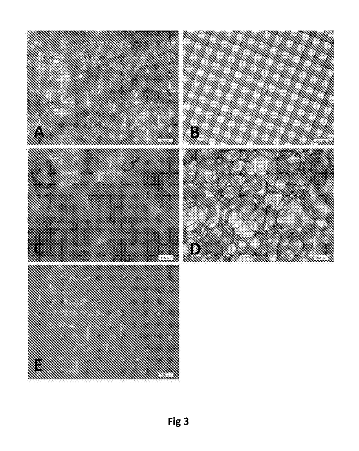
[0266]Placental ASCs were prepared as previously described. Initial seeding in tissue culture plate surfaces (“TCPS”) flasks and packed bed spinners with Fibracel disks was as follows. Briefly, ASCs (passage 4 cells) at 5-10×106 were thawed and ASC were plated at 1.5×10⁶ in each of 3 triple flasks with full DMEM. After 3-4 days post first seeding (-70% confluence) the cells were harvested and used to seed 5 triple flasks. Three to four days following the second seeding (˜70% confluence), cells were harvested and seeded four spinners with 8.1×10⁶ ASC each and 4 175 cm2 flasks with 0.5×10⁶ ASC each. Three days after the third seeding, the four flasks were harvested and seeded into 8 flasks (175 cm2) with 0.5×10⁶ ASC each.

TCPS Flasks

[0267]After 3 additional days, i.e., once cells in flask reached 70-80% confluence two flasks were used for RNA collection, two flasks for cell lysate, and two flasks for collectio...

example 2

Selection of Genes and Proteins for Differential Analysis

[0276]A panel of genes and proteins that are potentially differentially regulated in placental ASC-3D vs -2D cells was determined based on consideration of relevant biological pathways. The screening panels are presented in Tables 1-15, above.

example 3

Analysis of Proteins Secreted into Culture Medium and in Cell Lysates

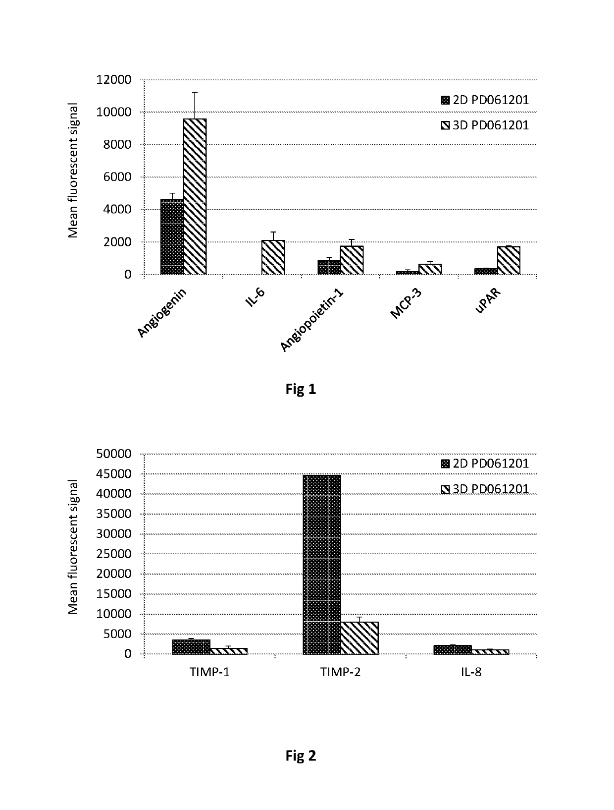
[0277]Conditioned medium and cell lysates were prepared as described in Example 1 was analyzed using RayBio® Human Angiogenesis and Human Inflammation Arrays (G series) (RayBiotech, Inc.) arrays according to the manufacture's instructions. Fluorescence was detected using a laser scanner and normalized, also according to the manufacture's instructions.

[0278]Tables 16 and 17 summarize the results of this experiment.

[0279]

TABLE 16Proteins Up-regulated in 3D Culture MediumDataPD280311PD061210t-TestsProteinCM 2DCM 3DCM 2DCM 3DPD280311PD061210Angiogenin425.54270.48555.3NA0.0020492233.64425.58547.84189.75055.910860808.223938.94803.510846.4AVG808.222696.954625.099594.01St DevNA1933.79391.901620.78IL-64820.801888.80.0004794819.201651.687.45971.602710.6257.65971.702284.2AVG172.565395.8502110.07St DevNA814.300510.98Angiopoietin 1622.55814.51013.315250.6021760.0041046651.1814.2991.41428.545.351213.4705.42217.5173.91128862.4190...


PUM


Abstract

Adherent stromal cells cultured under three dimensional conditions are provided, characterized, and distinguished from adherent stromal cells cultured under two dimensional conditions.

Description

[0001]This Application claims the benefit of U.S. Provisional Application No. 61 / 766,717, filed on Feb. 20, 2013, which is incorporated herein by reference in its entireity.INTRODUCTION[0002]In recent years, considerable activity has focused on the therapeutic potential of mesenchymal stromal cells for various medical applications including tissue repair of damaged organs such as the brain, heart, bone and liver and in support of bone marrow transplantations. Mesenchymal stromal cells are a heterogeneous population of adherent cells obtained from e.g. bone marrow, adipose tissue, placenta, and blood, that are capable of differentiating into different types of mesenchymal mature cells (e.g. reticular endothelial cells, fibroblasts, adipocytes, osteogenic precursor cells) depending upon influences from various bioactive factors. They have been widely studied in regenerative medicine as the foundation to build new tissues such as bone, cartilage and fat for the repair of injury or repl...

Claims


Application Information

Patent Timeline
Patent Type & Authority Patents(United States)
IPC IPC(8): C12Q1/68C12Q1/6881C12N5/0775C12N5/077G01N33/68A61K35/00
CPCC12Q1/6881C12N5/0652C12N5/0668G01N33/6803A61K35/00C12N2513/00G01N2333/96494G01N2333/475G01N2333/5412G01N2333/5415G01N2333/56G01N2333/78C12Q2600/158
Inventor OFIR, RACHELABRAHAM, EYTAN
Owner PLURI BIOTECH LTD
Learn More