
Device for dynamic controlling of the radiation level for radiation-based, real-time, medical-imaging systems

a dynamic control and radiation level technology, applied in the field of dynamic control of the radiation level of radiation-based, real-time, medical-imaging systems, can solve the problemsaffecting the safety of the imaging environment of the operator, and causing the patient to suffer various radiation-based diseases, etc., to achieve the effect of reducing the service life of the fluoroscope, reducing the total amount of radiation received by the patient, and limiting the total amount of radiation

Active Publication Date: 2019-10-08
EKIN EFE OZEL SAGLIK HIZMETLERI MEDIKAL TEKNOLOJI AR GE GIDA INSAAT SAN TIC LTD STI
View PDF4 Cites 0 Cited by
  • Summary
  • Abstract
  • Description
  • Claims
  • Application Information

AI Technical Summary

Benefits of technology

[0022]The object of the invention is to propound a system that reduces and limits the total amount of radiation received by the patient by checking the total amount of radiation received by the patient through a control mechanism.
[0023]Another object of the invention is to create a safer imaging environment for the operator by limiting the amount of radiation received by the operator.
[0024]Another object of the invention is to propound a system that enables increasing the amount of radiation, and thus image quality, in cases that require high resolution.
[0025]Another object of the invention is to enable adjustment of the image quality at any time during the operation under the control of the operator, thus propounding a more effective system capable of swifter operation.
[0026]Another object of the invention is to propound a system where the radiation dose can be adjusted dynamically; thus it is capable of eliminating operation errors and defects that might arise due to low resolution.

Problems solved by technology

However, high amounts of radiation capable of providing high quality images are harmful to the patient, operator and technicians.
While the high amount of radiation used improves the image quality, it might lead to various radiation-based diseases in the patient and the operator, such as cancer.
On the other hand, because keeping the radiation amount at low levels reduces image quality, this might lower the performance of the operation applied by the operator.
In the prior art, devices capable of performing radiation based on real-time imaging, such as fluoroscopy and cine acquisition and suchlike, lack the means to effectively control the radiation amount during the process.
When initiating the operation, the radiation level is set by means of a controller and radiation is applied to the patient during the process without effectively controlling the level.
However, as the footswitch used in existing systems has no control function other than turning the device on and off, it is not suitable for applying differing radiation regimes according to the needs of different patients.
Furthermore, working with a fixed frame rate during the operation and imaging process causes not only the patient but also the operator performing the operation to receive high doses of radiation.
However, it is not practical for the operator to continuously modify the frame rate applied during the taking of each film, thereby interrupting the applied process, and thus is not suitable.
Some methods are contemplated in the prior art for reducing and limiting the amount of radiation to the patient and the operator; however, these methods have proved to be insufficient.
However, this solution can only be employed under conditions where the device is used for imaging purposes but cannot be employed where the device is used for an imaging function assisting an operation.
However, this method enables involves measuring the amount of radiation dose received by the patient, and the imaging operation might be interrupted when the dose increases.
However, no structure associated with checking the radiation generated by the fluoroscope during surgery is disclosed within the system.
It is not possible to control the radiation received by the patient by use of this system.
However, if this application is used in any medical imaging system, it is not possible to protect the patient, as the amount of radiation received by the patient is not regulated.
However, the fact that the operator generates X-rays by activating the fluoroscope by looking causes the system to function slowly.
This increases overall operation time.
Another technical deficiency of the mentioned device is that continuous activation and deactivation of the x-ray source will damage the fluoroscope over time.
This reduces the service life of the fluoroscope.

Method used


Image

  • Device for dynamic controlling of the radiation level for radiation-based, real-time, medical-imaging systems

Examples


Embodiment Construction

[0001]The invention is a device for checking the amount of radiation emitted during the imaging process, particularly by devices capable of radiation-based real-time imaging, such as fluoroscopy and cine acquisition. The invention is particularly suitable for use by cardiologists and radiologists performing angiographic interventions in the field of medicine. The invention enables real-time controlling of the image quality and the image frame rate by means of a foot-switcher with the new function of being driven by a pressure-sensitive controller.

PRIOR ART

[0002]Fluoroscopy and cine acquisition are angiographic medical imaging techniques used for obtaining real-time radiological images of the patient.

[0003]In its simple form, a fluoroscope comprises an X-ray source and fluorescent display between which the patient is placed. In modern fluoroscopes, the display is connected to an X-ray concentrator and a video camera that enables monitoring or recording of the images on the display. T...


PUM


Abstract

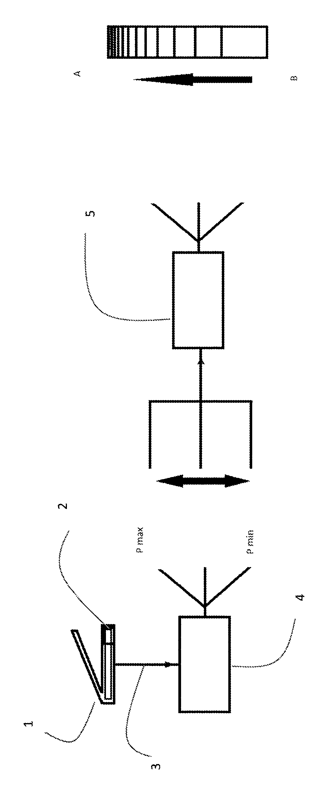
The invention is a device for controlling the amount of radiation emitted during the process of imaging, particularly by instruments capable of radiation-based real-time imaging, such as fluoroscopy and cine acquisition. The related device comprises a pressure-sensitive foot controller (1), a pressure sensor (2), a signal transmitter (3), a signal converter (4), and signal processing software (5). The invention enables real-time controlling of the image quality and the image frame rate by means of a foot-switcher with the new function of being driven by a pressure-sensitive controller.

Description

DESCRIPTION OF THE INVENTION[0001]The invention is a device for checking the amount of radiation emitted during the imaging process, particularly by devices capable of radiation-based real-time imaging, such as fluoroscopy and cine acquisition. The invention is particularly suitable for use by cardiologists and radiologists performing angiographic interventions in the field of medicine. The invention enables real-time controlling of the image quality and the image frame rate by means of a foot-switcher with the new function of being driven by a pressure-sensitive controller.PRIOR ART[0002]Fluoroscopy and cine acquisition are angiographic medical imaging techniques used for obtaining real-time radiological images of the patient.[0003]In its simple form, a fluoroscope comprises an X-ray source and fluorescent display between which the patient is placed. In modern fluoroscopes, the display is connected to an X-ray concentrator and a video camera that enables monitoring or recording of ...

Claims


Application Information

Patent Timeline
Patent Type & Authority Patents(United States)
IPC IPC(8): A61B6/00H05G1/56H05G1/30
CPCH05G1/56A61B6/542A61B6/467H05G1/30
Inventor ERCAN, ERTUGRUL
Owner EKIN EFE OZEL SAGLIK HIZMETLERI MEDIKAL TEKNOLOJI AR GE GIDA INSAAT SAN TIC LTD STI
Learn More