Automatic three-dimensional segmentation method for OCT and doppler OCT angiography

a three-dimensional segmentation and angiography technology, applied in the field of oct angiography, can solve the problems of limited clinical applications, risk of complications, and limited clinical applications, and achieve the effect of early diagnosis and precise monitoring

Active Publication Date: 2019-11-05
RGT UNIV OF CALIFORNIA
View PDF12 Cites 2 Cited by
  • Summary
  • Abstract
  • Description
  • Claims
  • Application Information

AI Technical Summary

Benefits of technology

[0009]In the invention, we combine the three dimensional dynamic programming method with intensity-based Doppler variance based on swept source OCT (SS-OCT). The microvascular morphology of intra-retinal layers are visualized in a normal subject by using this method. Imaging of the microvascular network with three dimensional structure of the retina has potential for earlier diagnosis and precise monitoring in retinal vascular diseases.
[0010]The illustrated embodiment of the invention includes a three-dimensional segmentation method with a Doppler variance method to visualize the microvascular network of the individual layers in retina. The microvascular network is obtained in a non-invasive way. The morphology of the microvascular network for individual layers can provide the clinicians valuable information for earlier diagnosis and precise monitoring in retinal vascular diseases.
[0011]The three-dimensional structure of retina is obtained by using a swept source optical coherence tomography (OCT) system. Each tomographic image is composed of eight repeat scans at the same position, which achieves a high time difference to improve the sensitivity of the angiographic method. A subpixel registration algorithm is used to reduce the eye movement. A graphics processing unit (GPU) was used to accelerate the data processing to achieve real-time preview of the acquired data.

Problems solved by technology

However, FFA is an invasive procedure with an associated risk of complications which limits clinical applications.
In addition, FFA can only provide two-dimensional images of the fundus, and the deeper capillary network may be not visualized well by FFA.
However, the minimum cut graph algorithm is computationally expensive which usually requires minutes to complete a three dimensional data segmentation.

Method used


Image

  • Automatic three-dimensional segmentation method for OCT and doppler OCT angiography
  • Automatic three-dimensional segmentation method for OCT and doppler OCT angiography
  • Automatic three-dimensional segmentation method for OCT and doppler OCT angiography

Examples


Embodiment Construction

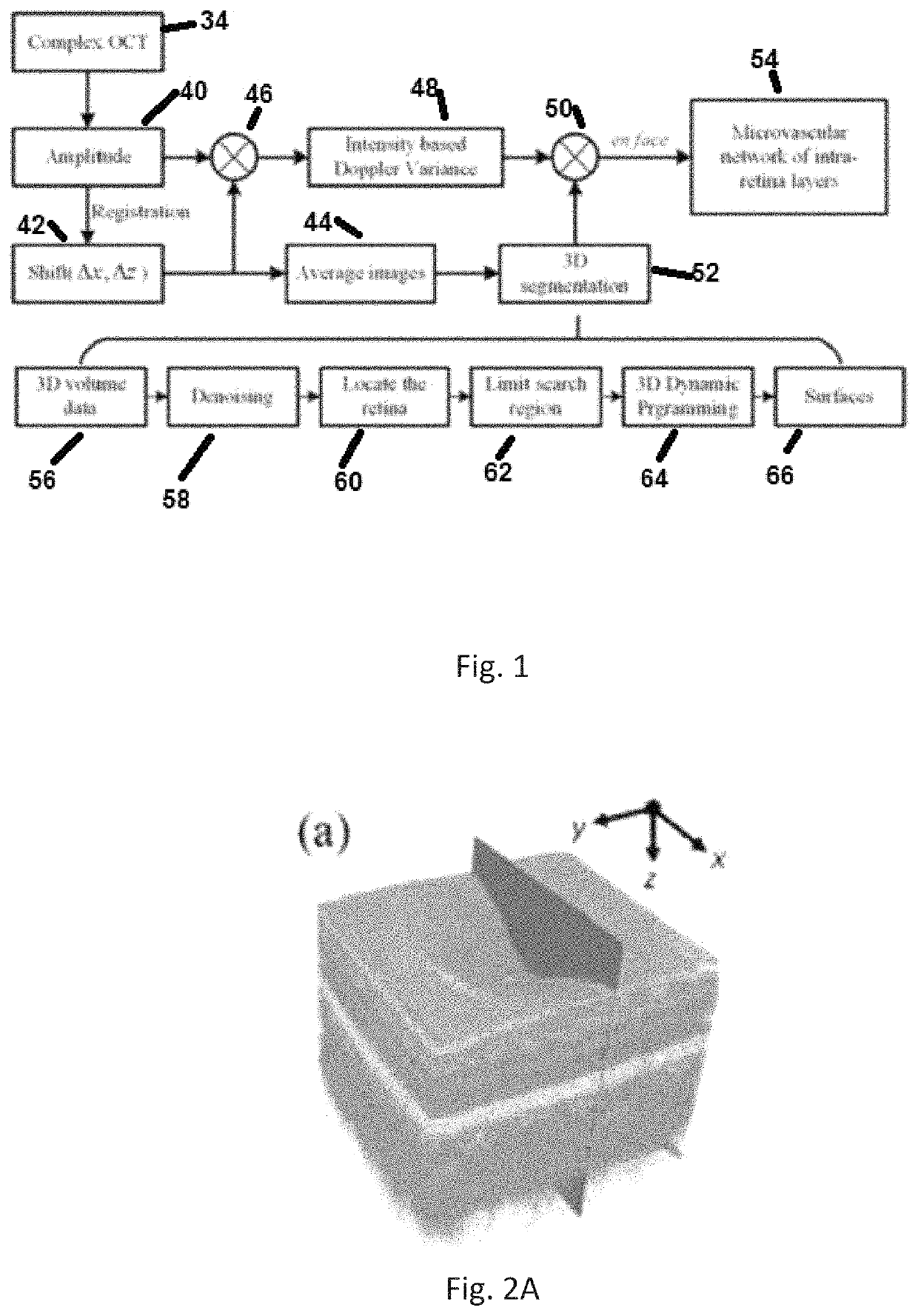
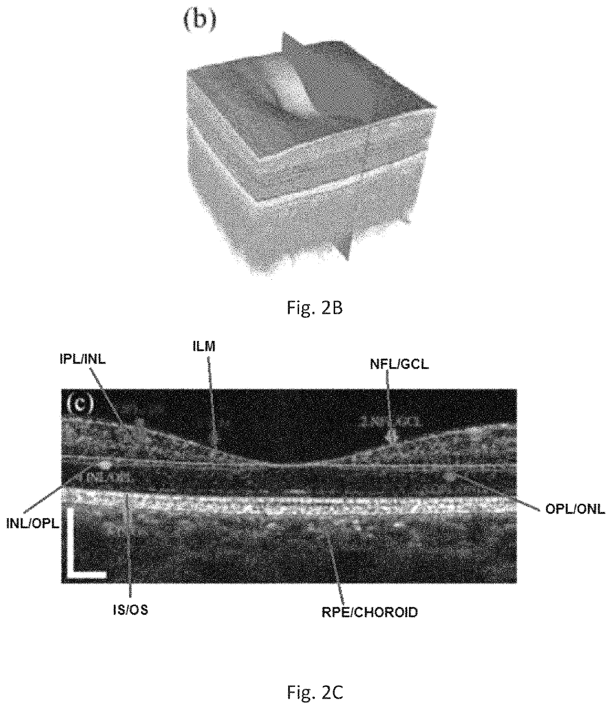
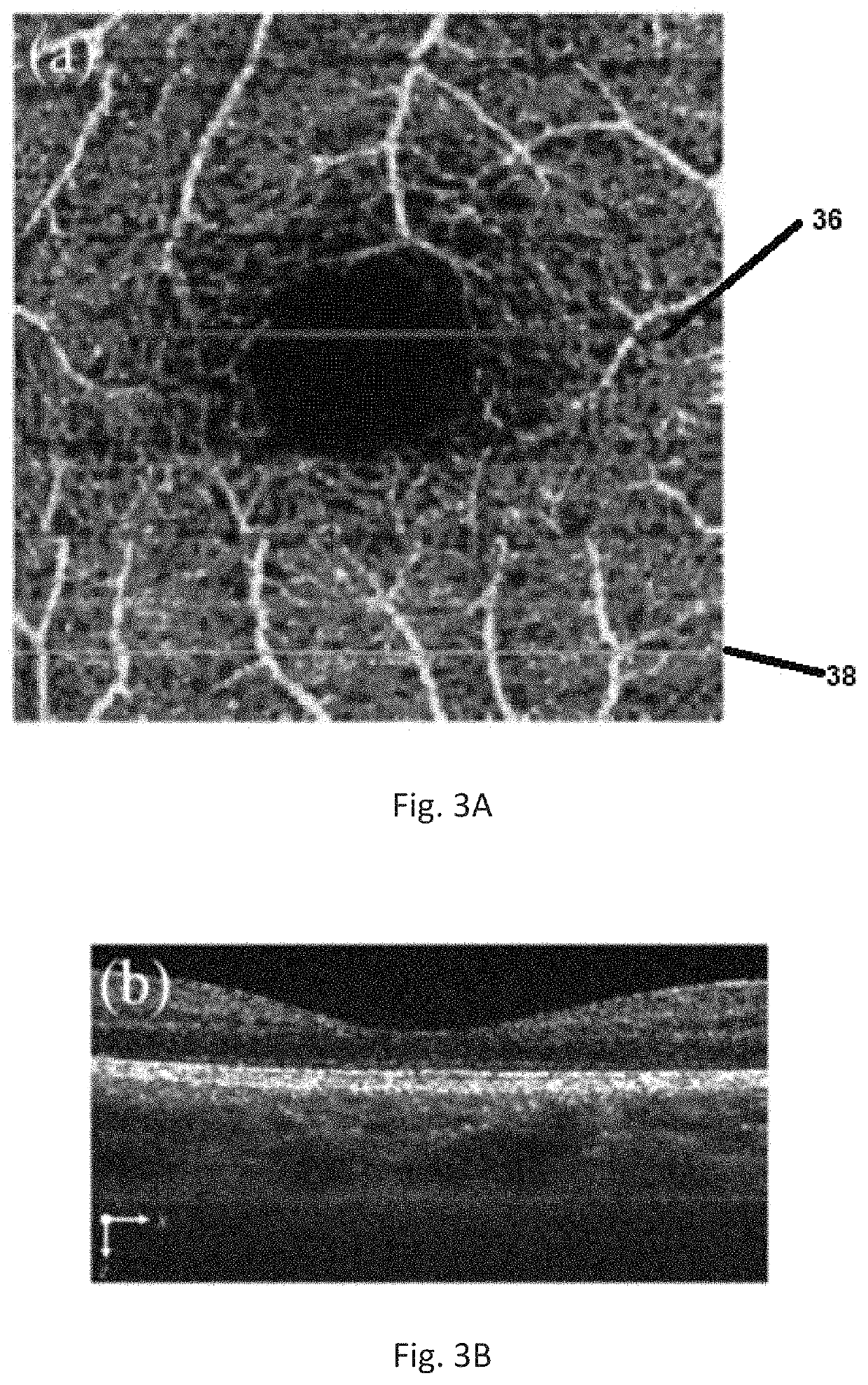
[0060]We have invented a functional optical imaging system for diagnosis of eye diseases. This system combines an automatic three-dimensional segmentation method with a Doppler variance method based on Fourier domain optical coherence tomography (OCT) to provide an earlier diagnosis and precise monitoring in retinal vascular diseases.

[0061]In the illustrated embodiment, a conventional 1050 nm swept-source OCT system 10 was used to acquire the three dimensional data of the fundus as diagrammatically shown in FIG. 5. A commercially available swept-source laser 12 (for example as are available from Axsun Technologies Inc., Billerica, Mass., USA) with a center wavelength of 1050 nm and 100 nm tuning range was used. The configuration of the system 10 is conventional and has already been described in previous publications, including “Ultrahigh speed 1050 nm swept source / Fourier domain OCT retinal and anterior segment imaging at 100,000 to 400,000 axial scans per second,” (Potsaid et al., ...


PUM

PropertyMeasurementUnit
depthaaaaaaaaaa
center wavelengthaaaaaaaaaa

Abstract

A three-dimensional (three dimensional) segmentation method with intensity-based Doppler variance (IBDV) based on swept-source OCT. The automatic three dimensional segmentation method is used to obtain seven surfaces of intra-retinal layers. The microvascular network of the retina, which is acquired by the IBDV method, can be divided into six layers. The microvascular network of the six individual layers are visualized, and the morphology and contrast images can be improved by using the segmentation method. This method has potential for earlier diagnosis and precise monitoring in retinal vascular diseases. Each tomographic image is composed of eight repeat scans at the same position, which achieves a high time difference to improve the sensitivity of the angiographic method. A subpixel registration algorithm is used to reduce the eye movement. The GPU was used to accelerate the data processing to achieve real-time preview of the acquired data.

Description

GOVERNMENT SUPPORT[0001]This invention was made with government support under contract numbers R91 EY-021529, R01HL-125084. P41-E6015890, funded by the National Institutes of Health. The government has certain rights in the invention. This invention was also made with support under contract number 2012YQ12008004, funded by the National Major Equipment Program from the Ministry of Science and Technology in Beijing, China. The government of China also has certain rights in the invention.RELATED APPLICATIONS[0002]This application is related to provisional patent application, entitled, Automatic Three-Dimensional Segmentation Method for OCT and Doppler OCT Angiography Ser. No. 62 / 183,072, filed on Jun. 22, 2015, and Ser. No. 62 / 183,601, filed on Jun. 23, 2015, under 35 USC 119, which are incorporated herein by reference.BACKGROUND[0003]Field of the Technology[0004]The invention relates to the field of OCT angiography, specifically an apparatus and method for an automatic three-dimension...

Claims


Application Information

Patent Timeline
Patent Type & Authority Patents(United States)
IPC IPC(8): A61B3/10A61B3/00G01N21/47
CPCG01N21/47A61B3/0025A61B3/102
Inventor CHEN, ZHONGPINGFAN, LUHUANG, SHENGHAI
Owner RGT UNIV OF CALIFORNIA
Why Eureka
Try Eureka