KIR3DL2 binding agents

a binding agent and kir3dl technology, applied in the field of kir3dl2 binding agents, can solve the problems of impeded adcc-based approaches and strong internalization of kir3dl2, and achieve the effect of avoiding competition with ligands

Active Publication Date: 2020-03-03
INNATE PHARMA SA
View PDF16 Cites 3 Cited by
  • Summary
  • Abstract
  • Description
  • Claims
  • Application Information

AI Technical Summary

Benefits of technology

[0009]We further provide anti-KIR3DL2 antibodies that are capable of causing an increase of cell surface KIR3DL2 polypeptide available for binding by an anti-KIR3DL2 antibody, notably on malignant cells. The antibodies may, in one embodiment, increase the level of expression of KIR3DL2 polypeptides on the cell surface (e.g. of malignant cells). The antibodies may, in one embodiment, increase the amount or number of KIR3DL2 polypeptides on the cell surface available for binding by an anti-KIR3DL antibody. The antibodies may, in one embodiment, stabilize and / or cause accumulation of KIR3DL2 polypeptides present on the cell surface, e.g., they may decrease receptor cycling or internalization of KIR3DL2 polypeptides. Antibodies that increase cell surface KIR3DL2, e.g. on pathogenic CD4+ T cells, have increased potency because they permit a greater number of antibodies to be bound to a KIR3DL2-expressing cell (e.g. target cell, malignant cell). In one embodiment, provided is an isolated monoclonal antibody that binds a KIR3DL2 polypeptide on the surface of a KIR3DL2-expressing cell, wherein the antibody causes an increase of the amount or numbers of KIR3DL2 polypeptides detectable at the cell surface after being in contact with cells (in vivo or in vitro) for at least 1 hour, 3 hours, 6 hours, 12 hours or 24 hours. The increase can be in comparison to a control antibody, e.g. an isotype control, or another antibody that binds KIR3DL2 (e.g. an antibody that has a different heavy and / or light chain variable region amino acid sequence).
[0013]In one aspect, the antibodies furthermore have the additional advantage of binding to multiple alleles of human KIR3DL2 while maintaining KIR3DL2 specificity over KIR3DL1.
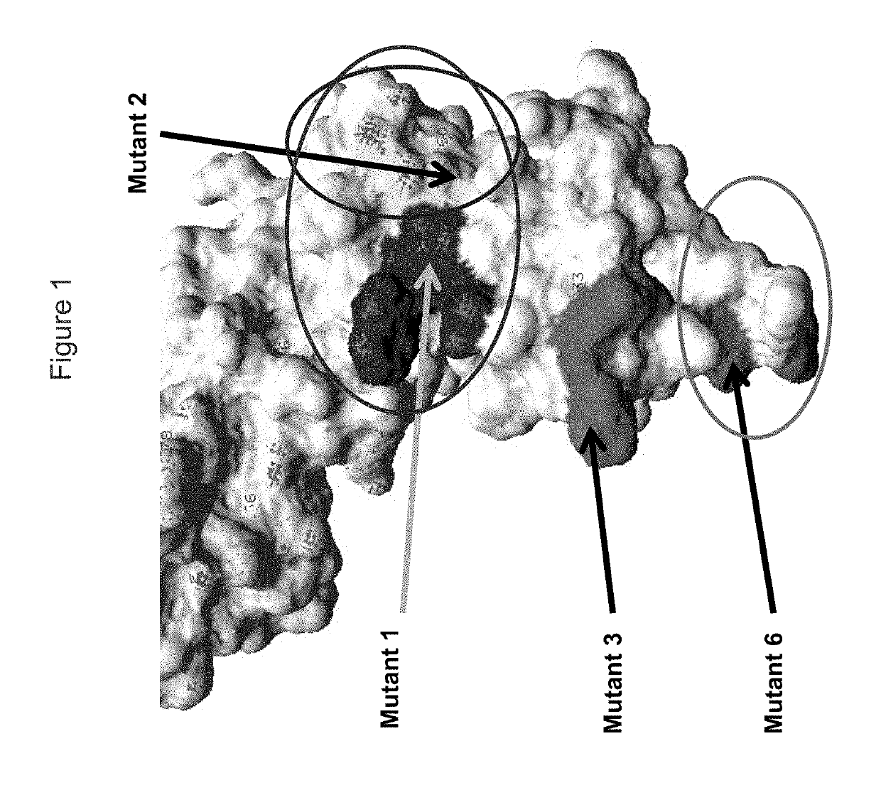
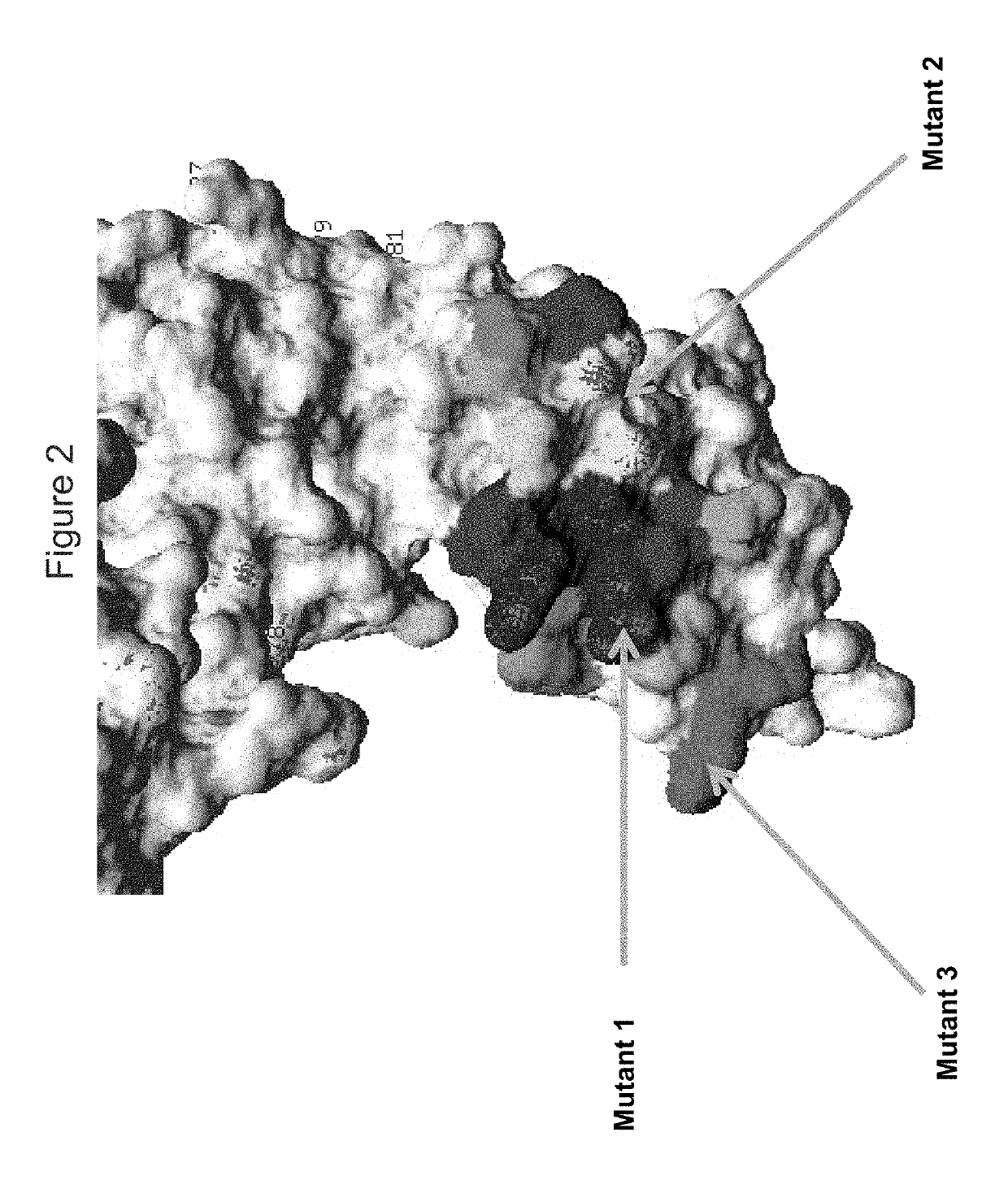
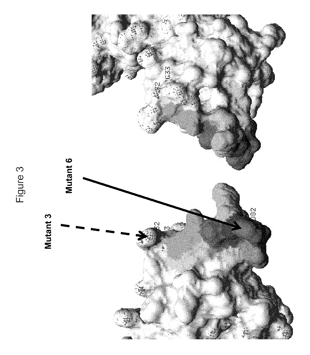
[0025]Preferably, in any of the embodiments herein, provided is an antibody binds to an amino acid residue within the D0 domain (residues 1 to 98 of SEQ ID NO: 1) and / or the D2 domain (residues 193 to 292 of SEQ ID NO: 1) of a KIR3DL2 polypeptide. Optionally, binding of the antibody to a KIR3DL2 polypeptide having a mutation at a residue within the D0 and / or D2 domain is substantially reduced, in comparison to binding to a wild-type KIR3DL2 polypeptide of SEQ ID NO: 1.
[0040]In one embodiment, the antibody optionally detectably reduces binding between the KIR3DL2 and an HLA class I-ligand of KIR3DL2 (e.g. HLA-B27, HLA-A3, HLA-B7, HLA-B35 and / or HLA-A2).
[0072](j) has reduced binding to a KIR3DL2 polypeptide having a mutation at one or more of residues R13, P14, S15, H23, A25, Q27, H32, G33, I60, G62, R78, L82, W226, 1231 and / or R246 of a KIR3DL2 polypeptide.

Problems solved by technology

It is demonstrated that KIR3DL2 internalization strongly hampers ADCC-based approaches.

Method used


Image

  • KIR3DL2 binding agents
  • KIR3DL2 binding agents
  • KIR3DL2 binding agents

Examples


example 1

n of Anti-KIR3DL2 Antibodies

Materials and Methods

Primary and Secondary Flow Cytometry Screenings

[0379]Anti-KIR3DL2 mAbs were primarily screened in flow cytometry for binding to KIR3DL2-expressing Sézary cell lines (HUT78 and COU-L) and to KIR3DL2-transfected tumor cell lines (HEK-293T). Flow cytometry devices include: FACSarray (BD Biosciences, primary screen), FACSCanto II No. 1 and No. 2 (BD Biosciences) (secondary screens) and FC500 (Beckman Coulter) (secondary screens). The KIR3DL2+ and other tumor cell lines used included:[0380]HUT-78 (KIR3DL2 positive Sézary cell line) grown in complete IMDM;[0381]HEK-293T (human kidney cancer) / KIR3DL2 and HEK-293T / KIR3DL2 Domain 0-eGFP cell lines (grown in complete DMEM);[0382]COU-L (KIR3DL2 positive Sézary cell line) (grown in complete RPMI complemented with 10% human serum AB);[0383]HEK-293T / KIR3DL1 and HEK-293T / KIR3DL1-eGFP cell lines (grown in complete DMEM);[0384]B221 (B-lymphoblastoid, CD20 positive human cell line) / KIR3DL2 cell line (g...

example 2

s that do not Induce KIR3DL2 Internalization

[0395]Briefly, either no antibody or 20 μg / mL of an anti-KIR3DL2 domain 0 antibody, or antibody 10F6, 2B12, 18C6, 9E10, 10G5, 13H1, 4B5, 5H1, 1E2, 1C3 or 20E9 were incubated with fresh Sézary Syndrome cells from 5 different human donors, for 24 h at 37° C. Cells were then washed, fixed and permeabilized using IntraPrep permeabilization reagent from Beckman Coulter. Presence of KIR3DL2-bound 10F6, 2B12, 18C6, 9E10, 10G5, 13H1, 4B5, 5H1, 1E2, 1C3 or 20E9 Ab is revealed with a goat anti-mouse Ab, labelled with GAM-PE. Table 6 shows an example of an anti-KIR3DL2 domain 0 antibody 13H1, after 24 h incubation, respectively. Table 5 shows a strong decrease in fluorescence for 13H1 in each of the different donors, confirming that the binding of this antibody down-modulates the expression of KIR3DL2 on SS cells. Similar results were obtained for anti-D0 antibody 4B5 as well as a range of anti-D1 antibodies. Conversely, the anti-KIR3DL2 domain 0 or ...

example 3

s that do not Internalize into Sézary Syndrome Cell Line

[0398]Internalization of antibodies 10F6, 2B12, 18C6, 9E10, 10G5, 13H1, 4B5, 5H1, 1E2, 1C3 and 20E9, as well as antibody AZ158 (an anti-domain 0 mAb) and other anti-D1 antibodies were assessed by fluoro-microscopy using the HUT78 SS cell line.

Materials and Methods:

[0399]Hut-78 cells were incubated during 1H at 4° C. with 10 μg / ml of the different antibodies. After this incubation cells were either fixed (t=OH) or incubated for 2H at 37° C. Cells incubated for 2H were then fixed and stained. Antibodies were stained using goat anti-mouse antibodies coupled to Alexa594 (Invitrogen, A11032). LAMP-1 compartments were stained using rabbit anti-LAMP-1 antibodies (Abcam, ab24170) revealed by goat anti-rabbit polyclonal antibodies coupled to FITC (Abcam ab6717). Pictures were acquired using an Apotome device (Zeiss) and analyzed using the Axiovision software.

Results:

[0400]Anti-KIR3DL2 mAbs were visible in red while LAMP-1 compartments w...


PUM

PropertyMeasurementUnit
concentrationaaaaaaaaaa
pHaaaaaaaaaa
median timeaaaaaaaaaa

Abstract

The present invention relates to methods for the treatment of cancer and inflammatory disease using antibodies (e.g. monoclonal antibodies), antibody fragments, and derivatives thereof that specifically bind KIR3DL2. The invention also relates to antibodies, cells producing such antibodies; methods of making such antibodies; fragments, variants, and derivatives of the antibodies; pharmaceutical compositions comprising the same.

Description

CROSS-REFERENCE TO RELATED APPLICATIONS[0001]This application is a continuation-in-part of International Application No.PCT / EP2013 / 069302 filed Sep. 17, 2013, which claims the benefit of U.S. Provisional Application No. 61 / 702,834, filed Sep. 19, 2012, the disclosures of which are incorporated herein by reference in their entireties; including any drawings.FIELD OF THE INVENTION[0002]The present invention provides antigen-binding proteins capable of binding to KIR3DL2 polypeptides. The antibodies have increased activity in the treatment of disorders characterized by KIR3DL2-expressing cells, particularly CD4+ T cells, including malignancies such as Mycosis Fungoides and Sézary Syndrome, and KIR3DL2-expressing autoimmune disorders.REFERENCE TO SEQUENCE LISTING[0003]The Sequence Listing for this application is labeled “Seq-List-replace.txt” which was created on Dec. 26, 2017 and is 91 KB. The entire content of the sequence listing is incorporated herein by reference in its entirety.BA...

Claims


Application Information

Patent Timeline
Patent Type & Authority Patents(United States)
IPC IPC(8): C07K16/28G01N33/569A61K39/00
CPCC07K16/2803G01N33/56966G01N33/56972C07K2317/732C07K2317/34G01N2333/70503C07K2317/76A61K2039/505C07K2317/565G01N2800/7095C07K2317/33C07K2317/77G01N2800/24C07K2317/734G01N2800/52A61P29/00A61P35/00A61P37/02A61K39/395
Inventor GAUTHIER, LAURENTKOLLNBERGER, SIMONROSSI, BENJAMINSICARD, HÉLÈNEPATUREL, CARINECORNEN, STÉPHANIEZERBIB, STÉPHANIE
Owner INNATE PHARMA SA
Why Eureka
Try Eureka