

Methods, systems, and compositions for determining blood clot formation, and uses thereof
- Summary
- Abstract
- Description
- Claims
- Application Information
AI Technical Summary
Benefits of technology
Problems solved by technology
Method used
Image

Examples
embodiments a1-a9
A. Embodiments A1-A9
Embodiment A1
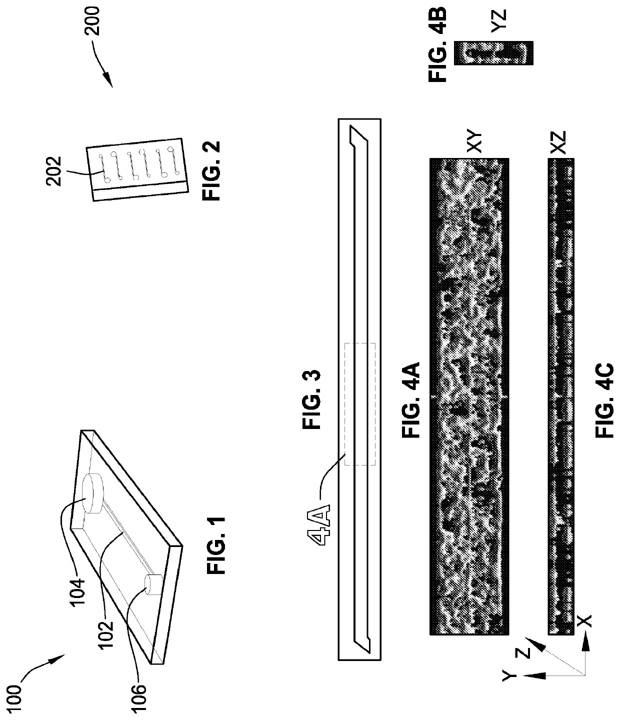
[0425]A microchannel comprising one or more surfaces, the microchannel having living endothelial cells on all of the microchannel surfaces.
Embodiment A2
[0426]The microchannel of embodiment A1, wherein the living endothelial cells are human umbilical vein endothelial cells.
Embodiment A3
[0427]The microchannel of embodiment A1, wherein the surfaces are coated with at least one attachment molecule that supports adhesion of the living endothelial cells.
Embodiment A4
[0428]The microchannel of embodiment A1, wherein the microchannel includes a top surface, a bottom surface, a first side surface, and a second side surface.
Embodiment A5
[0429]The microchannel of embodiment A4, wherein the bottom surface includes a membrane.
Embodiment A6
[0430]The microchannel of embodiment A1, wherein the microchannel is in fluid communication with an input port and an output port.
Embodiment A7
[0431]The microchannel of embodiment A1, wherein the microchannel has a width in the r...
embodiments d1-d8
D. Embodiments D1-D8
Embodiment D1
[0442]A method comprising:
[0443]1) providing[0444]a) a microchannel with one or more surfaces, and[0445]b) living endothelial cells on all of the surfaces; and
[0446]2) introducing fluid into the microchannel.
Embodiment D2
[0447]The method of embodiment D1, wherein the living endothelial cells are human umbilical vein endothelial cells.
Embodiment D3
[0448]The method of embodiment D1, wherein the fluid is selected from a group consisting of a blood sample, a serum sample, a plasma sample, a lipid solution, a nutrient medium, or a combination of two or more thereof.
Embodiment D4
[0449]The method of embodiment D1, wherein the fluid includes whole blood that contacts the endothelial cells without clotting.
Embodiment D5
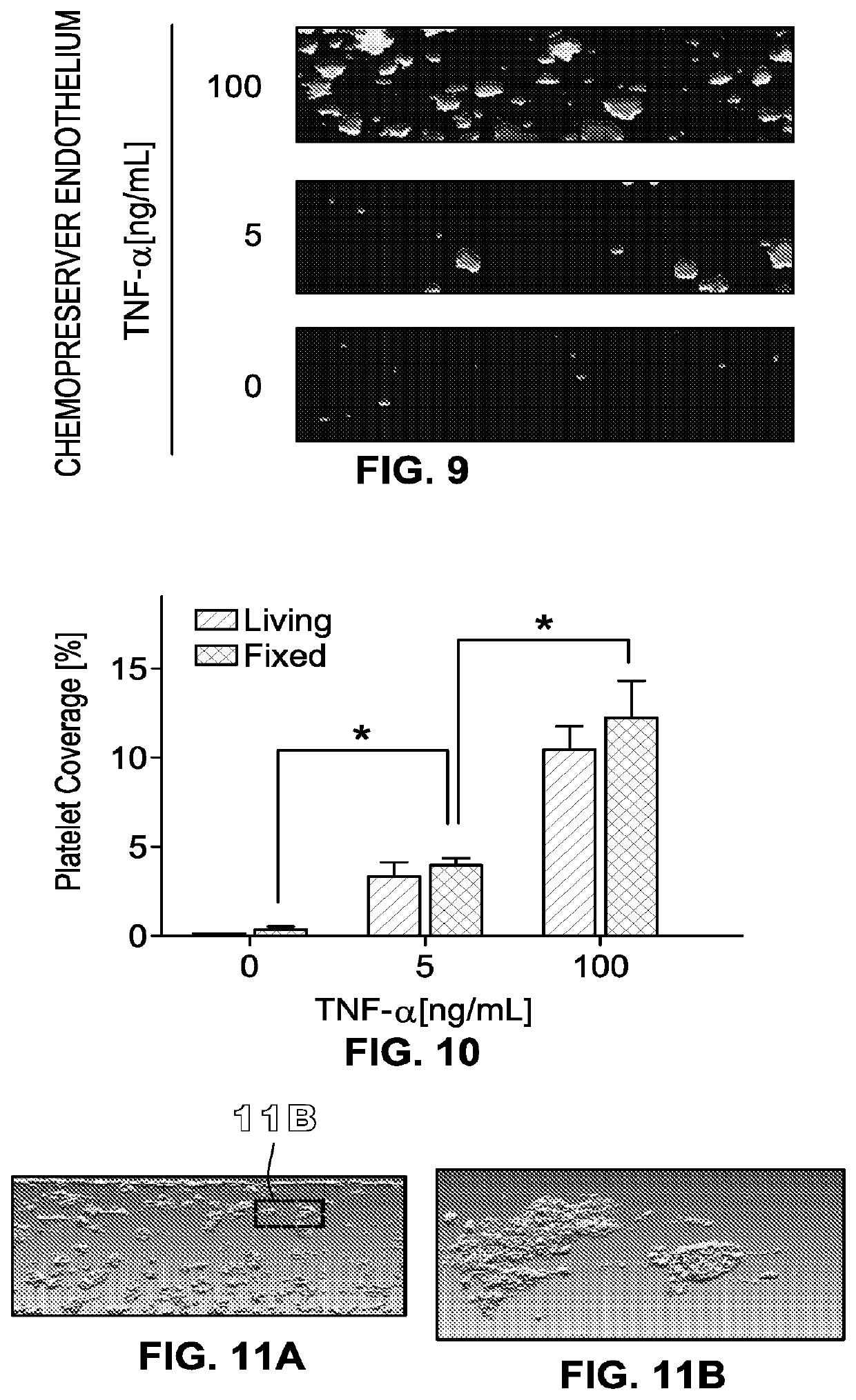
[0450]The method of embodiment D1, wherein the fluid includes platelets, the platelets contacting the endothelial cells without clotting.
Embodiment D6
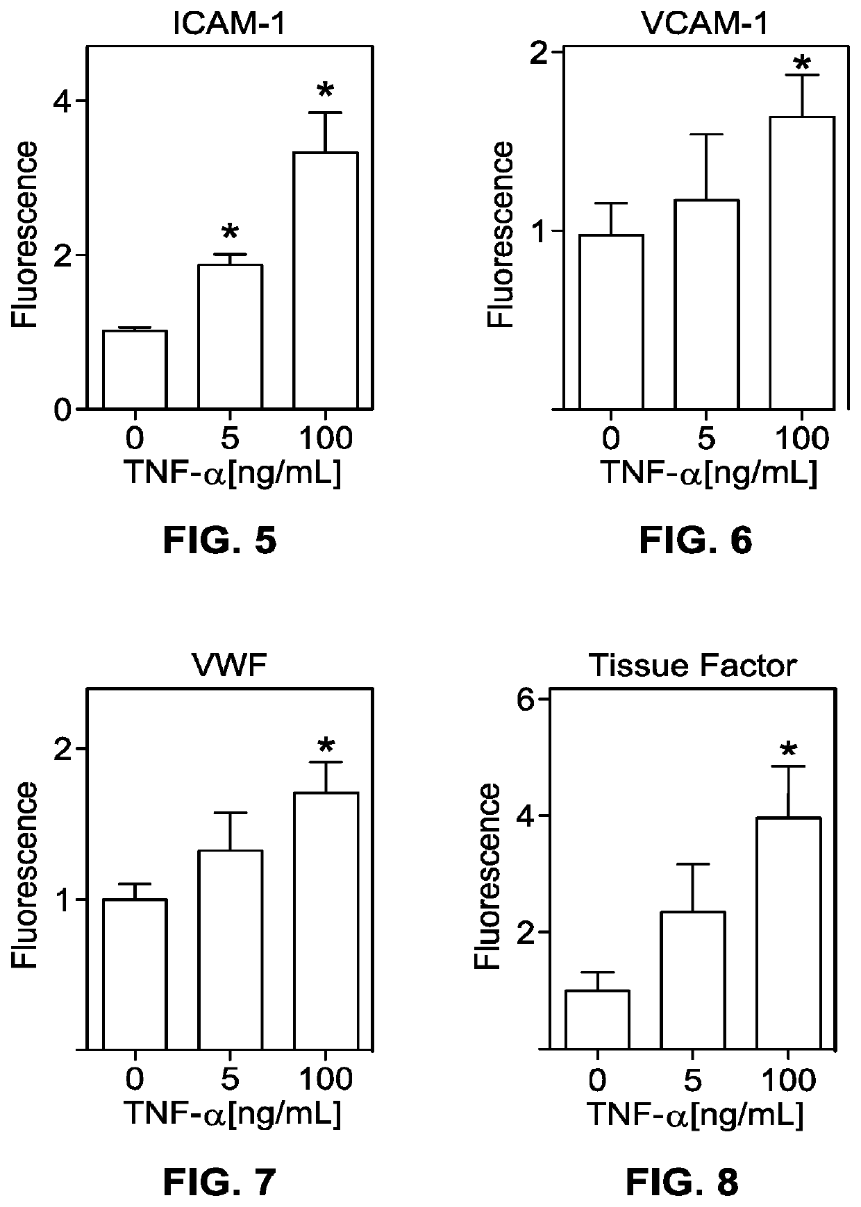
[0451]The method of embodiment D1, further comprising, prior to step 2), exposing the living e...
embodiment d8
[0453]The method of embodiment D6, wherein the fluid includes platelets, the platelets clotting upon contacting the endothelial cells.
PUM
 Abstract
Description
Claims
Application Information